Friday, April 30, 2010

Brain Tumor Awareness Month Fundraiser

-
May is Brain Tumor Awareness Month, and Channing Hall Charter School is fundraising for the cause.
Tomas Hollenbach, an 8-year-old student at the school, was diagnosed in February with an inoperable tumor on his brain stem. After going through 6 weeks of treatment, his tumor has shrunk but Hollenbach and his family are burdened with many medical expenses that Channing Hall is working to help with, using funds donated from those who appreciate the Hollenbach and his parents' strength throughout the ordeal.
There are a few of things the school is doing to raise money for the family:
Inspire Hope, Take Action: Support Brain Tumor Awareness Month. T-shirts emblazoned with this phrase will be sold at Channing Hall beginning May 5.
Tomas Hollenbach Charitable Account at America First Bank. Anyone can make a donation to Account #9040718.
Finally, Channing Hall is holding a raffle and yard sale at the school on May 22nd. They're still looking for raffle items from local businesses, so if you or anyone you know would be willing to make a donation during National Brain Tumor Awareness Month, don't hesitate.
Most of us are lucky enough not to be affected by brain tumors. We don't even think about something like this happening until it does- which is exemplified by FUTURE Brain Cancer Institute closing its doors in 2008 because of lack of funding. It seems that Utahns may be especially oblivious to this issue affecting over 600,000 people in the U.S., as we currently have no fundraising efforts for Brain Tumor Awareness, while there are annual walks in places like New York, San Francisco, even Delaware.
Appreciate how lucky you are to be unaffected by brain tumors- support Tomas Hollenbach and his family, or even go big for the cause as a whole and organize a walk by visiting the National Brain Tumor Society.

Brain Research to Ameliorate Impaired Neurodevelopment: Home-based Intervention Trial (BRAIN-HIT)

This randomized controlled trial aims to evaluate the effects of an early developmental intervention program on the development of young children in low- and low-middle-income countries who are at risk for neurodevelopmental disability because of birth asphyxia. A group of children without perinatal complications are evaluated in the same protocol to compare the effects of early developmental intervention in healthy infants in the same communities.

Birth asphyxia is the leading specific cause of neonatal mortality in low- and low-middle-income countries and is also the main cause of neonatal and long-term morbidity including mental retardation, cerebral palsy, and other neurodevelopmental disorders. Mortality and morbidity from birth asphyxia disproportionately affect more infants in low- and low-middle-income countries, particularly those from the lowest socioeconomic groups.

There is evidence that relatively inexpensive programs of early developmental intervention, delivered during home visit by parent trainers, are capable of improving neurodevelopment in infants following brain insult due to birth asphyxia.

Methods: This trial is a block-randomized controlled trial that has enrolled 174 children with birth asphyxia and 257 without perinatal complications, comparing early developmental intervention plus health and safety counseling to the control intervention receiving health and safety counseling only, in sites in India, Pakistan, and Zambia. The interventions are delivered in home visits every two weeks by parent trainers from 2 weeks after birth until age 36 months.

The primary outcome of the trial is cognitive development, and secondary outcomes include social-emotional and motor development. Child, parent, and family characteristics and number of home visits completed are evaluated as moderating factors.DiscussionThe trial is supervised by a trial steering committee, and an independent data monitoring committee monitors the trial.

Findings from this trial have the potential to inform about strategies for reducing neurodevelopmental disabilities in at-risk young children in low and middle income countries.Trial Registration: Clinicaltrials.gov NCT00639184

Author: Jan WallanderElizabeth McClureFred BiasiniShivaprasad GoudarOmrana PashaElwyn ChombaDarlene ShearerLinda WrightVanessa ThorstenHrishikesh ChakrabortySangappa DhadedNiranjana MahantshettiRoopa BelladZahid AbbasiWaldemar CarloBRAIN-HIT Investigators

If this is your brain on drugs, we want no part of drugs


illegaldrugs.jpg
We all remember those "This is your brain on drugs" ads, right? Featured a frying egg? Did that ever convince anyone to not use drugs? Yeah, we didn't think so. Today's video though? If the Partnership for a Drug Free America had this, they might have been able to claim "mission accomplished" years ago. What we have here is three nerdy dudes in skintight (and we mean skintight) body stockings. One red, one green, one blue (that's the band name: Red and Blue and Green). They are sporting some serious moose knuckles (that's the dude version of camel toe, fyi) and sort of spazzing out to some weird, repetitive electro pop in various locations -- laundromats, scenic vistas, recliners. All the while, a spoken/half-chanted of mantra illegal drugs is repeated ad infinitum.
Anyway, it's a disturbing picture of what "Illegal Drugs" might be like. If we were shown this as part of our DARE propaganda, we can't help but think we would have steered far clear of drugs -- not just illegal, but legal as well. Can't be too careful after seeing something like this.


Scientists race to create cat-level artificial brain

Complex task akin to reverse-engineering biological design of the brainPentagon-backed scientists aim to create a human-like machine, at some point. But they are starting out with the goal of crafting artificial intelligence on the level of a cat's brain. Still there are vast challenges.
If they get far enough, however, one scientist says that they could theoretically achieve feline intelligence with a mouse-sized artificial brain and an even smaller body.
That's because bigger brains by themselves don't necessarily mean greater intelligence or more complex behavior — for instance, cats show more smarts than cows despite having a feline brain 10 times smaller than a bovine brain. What might really matter is that humans and some other species have bigger brains for their body size. 
"If they're trying to get cat-level behavior, there's no necessary reason to go for a cat-level brain unless they need a cat-level body of some kind," said Mark Changizi, a neurobiologist at Rensselaer Polytechnic Institute in Troy, N.Y.
Changizi discussed his idea with an IBM researcher working on the SyNAPSE project run by the U.S. Defense Advanced Research Projects Agency (DARPA). The Pentagon agency has recruited the help of IBM, HP and leading research institutes to try to develop an artificial brain that is approximately cat-like in terms of size, number of brain cells and synapses, anatomical structure and even behavioral complexity.
Such a massive and complicated undertaking could easily falter short of the goal, even if various researchers have begun working on electronic devices that can mimic cat brain cells. Changizi compared the task at hand with trying to reverse-engineer the biological design of the brain and trace its evolution backwards over hundreds or thousands of years.
A mystery of bigger brains
But reservations aside, Changizi does see the small ray of hope for the DARPA project regarding the brain size required to achieve feline intelligence. His observation goes to the heart of what he calls the "big embarrassment of neurobiology," or the scientific uncertainty about why brain sizes scale up
so much in bigger bodies.
"Animals which are a thousand times bigger are just as dumb as the ones that are small," Changizi told LiveScience.
Bigger brains do allow for quantitative rather than qualitative improvements, such as finer resolution, higher sensitivity or greater precision in certain senses. They pack in more neurons, and add even more synapses (connections with other neurons) per neuron to keep all of the brain cells interconnected so that they can send signals to one another. 
Bigger brains also tend to have more compartments, where well-connected regions of the brain are located physically close together as a way of minimizing wire costs and neural delay between neurons. But greater intelligence does not seem to depend on having more compartments, more complex wiring or more neurons.
To better appreciate the puzzlement among neuroscientists, consider that a large mammal's brain is about a million times bigger than an insect's brain. Yet a survey of behavioral studies showed that mammals only have about two or three times as many behavioral functions compared with insects. Complex social behaviors among ants, bees and other insects also suggest their tiny brains can still pack a lot of behavioral complexity.
Artificial neural networks have even demonstrated that a relatively few neurons can carry out fairly complex cognitive tasks. Insects would likely have evolved over millions of generations to maximize their tiny brains' computing power — something that human scientists have only recently begun trying to recreate.
Boost the brain, shrink the body
Size only matters when an organism has a high brain-body size ratio. In other words, certain species such as humans have relatively big brains for their body size, as compared with the brain-body size ratios of other species. Mammals in particular show a higher number of behaviors when they have bigger brains relative to their body sizes.

Even if neuroscientists don't currently understand the connection with brain-body size ratio, Changizi says that solving the mystery might make the DARPA effort a bit easier. Rather than painstakingly recreating an artificial cat brain, researchers could make smaller artificial brains with even smaller bodies that are still capable of carrying out complex behaviors or tasks.
Changizi speculated that smarter brains might depend upon the diversity of neuron types, or a more efficient division of labor within the mind. But he readily acknowledged that most of the brain remains a mysterious "black box" for scientists.
"Lay people tend to think that we neuroscientists know what we're doing and that we're on the cusp of understanding it all, but that's so far from the truth," Changizi said.

The Talents of a Middle-Aged Brain

After we hit 40, many of us begin to worry about our aging brains. Will we spend our middle years searching for car keys and forgetting names?
The new book “The Secret Life of the Grown-Up Brain: The Surprising Talents of the Middle-Aged Mind,” by Barbara Strauch, has the answers, and the news is surprisingly upbeat. Sure, brains can get forgetful as they get old, but they can also get better with age, reports Ms. Strauch, who is also the health editor at The New York Times. Ms. Strauch, who previously tackled teenage brains in her book “The Primal Teen,” spoke with me this week about aging brains and the people who have them. Here’s our conversation:
Barbara Strauch
Q.   After exploring the teenage brain, why did you decide to write a book about grown-ups?
A.   Well, I have a middle-aged brain, for one thing. When I would go give talks about “The Primal Teen,” I’d be driven to the airport or back by a middle-aged person, and they’d turn to me and say: “You should do something about my brain. My brain is suddenly horrible. I can’t remember names.” That’s why I started looking into it. I had my own middle-aged issues like going into an elevator and seeing somebody and thinking, “Who are you?”
Q.   So what’s the bad news about the middle-aged brain?
A.   Obviously, there are issues with short-term memory. There are declines in processing speed and in neurotransmitters, the chemicals in our brain. But as it turns out, modern middle age is from 40 to 65. During this long time in the middle, if we’re relatively healthy our brains may have a few issues, but on balance they’re better than ever during that period.
Q.   Do teenage brains and middle-aged brains have much in common?
A.   The thing the middle-aged brain shares with the teenage brain is that it’s still developing. It’s not some static blob that is going inextricably downhill. Scientists found that when they watched the brains of teenagers, the brains were expanding and growing and cutting back and shaping themselves, even when the kids are 25 years old. I think for many years scientists just left it at that. They thought that from 25 on, we just get “stupider.” But that’s not true. They’ve found that during this period, the new modern middle age, we’re better at all sorts of things than we were at 20.
Q.   So what kinds of things does a middle-aged brain do better than a younger brain?
A.   Inductive reasoning and problem solving — the logical use of your brain and actually getting to solutions. We get the gist of an argument better. We’re better at sizing up a situation and reaching a creative solution. They found social expertise peaks in middle age. That’s basically sorting out the world: are you a good guy or a bad guy? Harvard has studied how people make financial judgments. It peaks, and we get the best at it in middle age.
Q.   Doesn’t that make sense, since our young adult lives are often marked by bad decisions?
A.   I think most of us think that while we make bad decisions in our 20s, we also have the idea that we were the sharpest we ever were when we were in college or graduate school. People think if I tried to go to engineering school or medical school now, I couldn’t do it. Because of these memory problems that happen in middle age, we tend to think of our brains as, on the whole, worse than in our 20s. But on the whole, they’re better.
Q.   So what’s happening in middle age that leads to these improvements?
A.   What we have by middle age is all sorts of connections and pathways that have been built up in our brain that help us. They know from studies that humans and animals do better if they have a little information about a situation before they encounter it. By middle age we’ve seen a lot. We’ve been there, done that. Our brains are primed to navigate the world better because they’ve been navigating the world better for longer.
There also are some other physical changes that they can see. We used to think we lost 30 percent of our brain cells as we age. But that’s not true. We keep them. That’s probably the most encouraging finding about the physical nature of our brain cells.
Q.   Is there anything you can do to keep your brain healthy and improve the deficits, like memory problems?
A.   There’s a lot of hype in this field in terms of brain improvement. I did set out to find out what actually works and what we know. What we do with our bodies has a huge impact on our brains. Our brains are more like our hearts in that everything you do for your heart is thought to be equally as good or better for your brain. Exercise is the best studied thing you can do to your brain. It increases brain volume, produces new baby brain cells in grownup brains. Even when our muscles contract, it produces growth chemicals. Using your body can help your brain.
Q.   What about activities like learning to play an instrument or learning a foreign language?
A.   The studies on this are slim. We’ve all been told to do crossword puzzles. Learning a foreign language, walking a different way to work, all that is an effort to make the brain work hard. And it’s true we need to make our brains work hard. One of the most intriguing findings is that if you talk to people who disagree with you, that helps your brain wake up and refine your arguments and shake up the cognitive egg, which is what you want to do.
Q.   Do social connections and relationships make a difference in how the brain ages?
A.   There is a whole bunch of science about being social and how cognitive function seems to be better if you are social. There is a fascinating study in Miami where they studied people who lived in apartments. Those who had balconies where they could see their neighbors actually aged better cognitively than others. There are a whole bunch of studies like that. People who volunteer and help kids seem to age better and help their brains. We forget how difficult it is to meet, greet and deal with another human being. It’s hard on our brains and good for them.
Q.   What was the most surprising thing you learned about the middle-aged brain?
A.   The hope I saw from real scientists was surprising. A lot of the myths we think of in terms of middle age, myths that I grew up with, turn out to be based on almost nothing. Things like the midlife crisis or the empty nest syndrome. We’re brought up to think we’ll enter middle age and it will be kind of gloomy. But as scientists look at real people, they find out the contrary. One study of men found that well-being peaked at age 65. Over and over they find that middle age, instead of being a time of depression and decline, is actually a time of being more optimistic overall.

Bret Michaels returns to hospital due to complications from his brain hemorrhage

a
"Health is a state of complete physical, mental and social well-being, and not merely the absence of disease or infirmity." ~World Health Organization, 1948

What does it take to be hospitalized for massive brain hemorrhaging and seizures one night and then be planning to tour only a little over a month later barring unforeseen complications? A complete devotion and passion for what you love. And that is what Bret Michael's, 47, showed us, until a complication arose, which led him back to the hospital. The mind/body connection, including attitude, desire, devotion, and passion are extremely powerful forces which are believed to aid in more speedy recoveries from physical ailments.

Bret Michael's was the lead singer for the '80s glam-metal "hair band" Poison, a judge on Nashville Star, a reality star headlining the VH1 show "Rock of Love with Bret Michaels", and just recently appeared on "Celebrity Apprentice" working hard to raise money for his charity, the non-profit organization, The American Diabetes Association.

Michael's suffered from a subarachnoid hemorrhage which is a type of bleeding stroke that occurs between the tissues around the brain and the brain itself. Sometimes there is simply no answer to what causes such strokes. Prognoses of this kind of complication depend on amount of bleeding, age, and symptom severity. About 35% of people die when they have a subarachnoid hemorrhage due to an aneurysm because it results in extensive brain damage. Another 15% die within a few weeks because of bleeding from a second rupture. People who survive for 6 months but who do not have surgery for the aneurysm have a 3% chance of another rupture each year.

Michael's responded well to his initial treatment; however, had another rupture and now remains in critical condition in ICU under 24 hour supervision by doctors and medical staff. Sources say that the next 7-10 days will be the most critical as an additional rupture or other complications may arise. We hope for Bret's strong mental attitude to continue to help with a speedy recovery.

Bret had to cancel some shows due to his hospitalization, but still plans to continue his tour on May 26, 2010.

Bret Michaels’ Doctors Hopeful for a ‘Full Recovery’

The rocker is in “good spirits” after the brain hemorrhage he suffered last week, but doctors say his recovery may take weeks or even months.
bret_michaels.pngBret Michaels’ family, friends and fans breathed a much-awaited sigh of relief when they learned that his condition is slowly improving after undergoing major brain surgery last week.
Doctors for the 47-year-old former Poison front man and top Celebrity Apprentice 3 contender said his recovery will be slow, but that he “is very lucky, as his condition could have been fatal,” his tour manager, Janna Elias told fans on his website, BretMichaels.com on April 27. Due to the severity of the hemorrhage, Michaels continues to remain hospitalized in an intensive care unit in critical but stable condition at an undisclosed hospital, she said.
Despite his ordeal, Michaels is conscious, talking slowly and in good spirits, his rep told People April 28.
Ten days after the Rock of Love star underwent an emergency appendectomy, he was rushed to the hospital again on April 22 with a headache he said felt like getting “hit with a baseball bat again and again,” a source told People earlier this week.
His massive headache came from a subarachnoid hemorrhage, a type of stroke that causes bleeding in the fluid-filled spaces around the base of the brain, wrote Elias. “It presents itself suddenly as the sound of a loud gunshot or thunderclap at the back of the head causing severe cranial pain and muscle spasms,” she said.
With further testing and rehabilitation, she said, doctors “are hopeful that Bret will gradually improve as the blood surrounding the brain dissolves and is reabsorbed into his system, which can be a very painful recovery and take several weeks to months.”
Michaels remains under 24-hour observation in the ICU, but “is in positive spirits,” she wrote. “He is responding well to tests and treatments.”
An Unsettling Complication
Even though Michaels’ condition has stabilized, he faces a new medical setback — seizures caused by a lack of sodium in the body, which is a common side effect from a brain hemorrhage, Elias wrote.
Still, she remains optimistic. “Even though today was a minor setback doctors remain hopeful for a full recovery and plan to release more specific information next Monday,” she said.
While some people are speculating that the stroke may have come after he was struck in the head by a falling piece of the set at the Tony awards last June, he will undergo further tests throughout the week to try to detect the exact cause of the rupture, said Elias. “Coupled with the fact that Michaels is a lifelong Type 1 diabetic and has recently undergone emergency appendectomy surgery while on tour in San Antonio, he will remain monitored closely by his medical team to make sure no complications occur from the diabetes.”
Friends Rally for Their Beloved Pal
Friends of Michaels from around the world have come forth to wish him well. Sharon Osbourne, who has been a friend of the singer’s for 30 years and stars with him on Apprentice, told US Magazine, “It’s devastating, and it makes you feel all these different things. Your heart goes out to his family, your heart goes out to him. He’s the nicest guy in the world. And then you start thinking about your own mortality. You start thinking, ‘Oh my God.’ It just makes you appreciate how lucky we all are.”
Apprentice honcho Donald Trump said on Today that Michaels “would work so hard, maybe harder than anybody else. And he’s doing well.”
Michaels’ longtime friend and guitar player in the Bret Michaels Band, Pete Evick, thanked fans on Michaels website for their outpouring of support, saying “we who are closest to Bret are overwhelmed with the amount of love and well wishes he is receiving. I know that everyone is waiting for some official news, however we just can’t give any right now. This is a time for family, a time for healing and a time for love.”
He praised Michaels for his work ethic, saying “I thought I was a hard worker. I didn’t understand that a man ten years older than me, battling a lifelong war with juvenile diabetes, more fame than most, and more money than he knows what to do with, could possibly get up every day with more drive and passion than myself. I was wrong! I didn’t understand what it meant to be a loyal friend. The kind that would do anything you ever needed without even asking why, what, who or where.
“Bret is the kind of guy that wakes up every day and treats it like it is his first, and may be his last,” he wrote. “He is full of passion, energy and love. But most of all, he is a survivor. Against the odds of a critical music industry, or battling a crippling disease, over and over again Bret has come out on top and ahead of the game. I personally expect nothing less from him in this situation.”
A New Michaels’ Song Released
To thank fans for their support, Evick released a “teaser” from Michael’s upcoming album, Custom Built, on the Web Page Audio portion of Michaels website. Evick released part of a song called “Wasted Time,” which he writes is “full of Bret Michaels’ emotion and heart. While not the final mix, I took some of the vocal we have laid down on the bus during the tour and comped together a piece of NEW MUSIC for you all. I believe all the fans will agree with me that this teaser is an amazing song. I hope you all use it, like I have this week, to remind you how amazing my great friend Bret is.”

Q: Is pregnancy brain a myth?

A: Probably. Research suggests pregnant women are no more forgetful than other women.
pregnancy_brain_300  
Have your say
What's been your experience of 'pregnancy brain'? Did you, or someone you know, have problems remembering things? Have your say on the messageboard below.
Conditions of Use
It's always been considered part and parcel of pregnancy, along with a swollen belly, varicose veins and food cravings. It' s so-called 'pregnancy brain' (sometimes called 'placenta brain' or 'mumnesia'); a pattern of woolly thinking and the forgetfulness that seems to accompany pregnancy.
Many pregnant women believe they experience it, which is not surprising given that most pregnancy books and manuals warn them to expect it.
Some people say 'pregnancy brain' is caused by high levels of oestrogen and other sex hormones during pregnancy – one theory even suggests it's an evolutionary development that allows a pregnant woman to become oblivious of outside distractions, so she can focus on the all-important task of preparing for the birth.
But is there any evidence for pregnancy brain?

No basis in fact

New research suggests not, says Professor Helen Christensen, director of the Centre for Mental Health Research at the Australian National University.
Often women think they have poor memory, yet objective tests show their memory is perfectly normal, she says.
Christensen and other Australian researchers concluded this after studying 1,241 women over an eight-year period. The researchers took a random sample of women from the electoral roll, aged 20 to 24 at the beginning of the study, and followed them up over time, watching to see who got pregnant. (Of the 1,241 women, 76 were pregnant when they had their first interview, 188 became mothers during the study – but were not pregnant during the first interview – and another 542 didn't get pregnant. The rest were dropped from the study for a variety of reasons).
All the women were given standard tests of mental ability to see how well they could recall and repeat words and numbers. The pregnant women were given tests before, during and after their pregnancies; and their results were compared to the test results of the women who didn't become pregnant.
The researchers found no essential differences between pregnant versus non-pregnant women. Some women in late pregnancy appeared to take longer to do a speed task, but the researchers say this effect might not be real and it needs to be further investigated before it can be confirmed.
Their overall conclusion was that 'pregnancy brain' has no basis in fact.

A myth explained

So why then do so many pregnant women believe they are foggy-brained?
Christensen thinks it may be that these women, especially in the later stages of pregnancy and after childbirth, simply have a lot on their mind. So what appears to be poor memory could be just a proneness to distraction, as the women ponder the tasks that lie ahead. (However, the study didn't actually test this hypothesis).
Also, some pregnant women are prone to anxiety and depression. Both these conditions can affect memory regardless of whether you are pregnant or not, Christensen says.
Another possible explanation is sleep deprivation – a common complaint throughout pregnancy and the early years of motherhood. Sleep deprivation is known to contribute to forgetfulness, in men as well as women.
And for some women, the expectation of poor memory may become self-fulfilling, says Christensen. These women believe pregnancy brain is a normal part of pregnancy, so they look for signs that they are experiencing it.
So women don't need to plan their nine-month odyssey on the expectation of a foggy mental state. That's the good news.
The bad news: pregnant women who don't want to be pestered may no longer have the excuse of pregnancy brain. Although they still have plenty of other excuses – fatigue, swollen feet, indigestion…
Dr Helen Christensen is the director of the Centre for Mental Health Research, at the Australian National University in Canberra. She spoke to Peter Lavelle.

'Cyborg brain op gives Barry new lease of life

Barry Salter with wife Sheila
A PARKINSON’S sufferer has told how a miracle operation has transformed his life – allowing him to ditch his wheelchair and become a healthy sportsman.

Grandfather Barry Salter, 61, was struck down with neurological disorder Parkinson’s at the age of 48 and could not wash, dress or feed himself.

He was confined to a wheelchair and had to down a cocktail of 20 different pills a day to combat crippling aches and pains and symptoms including hallucinations and anxiety.

But thanks to a revolutionary “brain pacemaker” fitted by surgeons in an eight-hour operation, Barry’s life has been turned around.

He now goes to the gym twice a week, practises martial arts, swims and plays table tennis regularly.

Barry has even been able to make two trips across the globe to visit friends in Australia, whom he thought he would never see again.

Delighted Barry compared himself to Star Trek creatures “the Borg” and said the Deep Brain Stimulator operation could transform the lives of thousands of fellow sufferers in the UK.

The former health service manager from Newmarket  said: “Parkinson’s is like an insidious disease, it creeps up on you and gets worse.

“My movement deteriorated, I wasn’t sleeping, I was anxious, suffering aches and pains and hallucinations and had to give up work.

“It had a devastating effect. It got so bad I had a wheelchair. I was only in my 50s.”

But neurologist Dr Graham Lennox, of Newmarket Hospital, provided a lifeline by recommending the Deep Brain Stimulator (DBS) operation.

During specialist surgery at the National Hospital for Neurology and Neurosurgery in London, four electrodes, in the form of 5in-long titanium rods, were placed inside his brain.

The brain pacemaker sends electrical impulses into the titanium electrodes and alleviates symptoms of Parkinson’s.

Now Barry carries a remote control to regulate how much voltage the pacemaker administers to his brain.

He said: “I feel like the Borg from Star Trek. Now I work out in the gym twice a week and do tai chi as well.”

The rods and wires mean Barry cannot walk through airport security scanners and was once quizzed by staff at Next, in Cambridge, after setting off the door alarms.

Barry said the operation in 2005 meant he could look forward to enjoying life again with his wife Sheila, 60.

Dr Keiran Breen, director of research and development at Parkinson’s UK, said the operation was clearly a success and should be made available to more sufferers.
 

Creativity, the Brain, and Evolution

Creativity: Adaptation or a byproduct of increased intelligence?

We humans are a creative species. When we compare what we do and make with what other species do and make, it is self-evident that we are the most creative species. But what has the role of creativity been over the course of our evolution? Is it just a by-product, an emergent property, of more fundamental cognitive processes, such as those involving problem-solving ability, memory, language, attention, and so on? Or is creativity a distinct and identifiable cognitive process of its own, which varies from individual-to-individual? Heritable genetic variation is the raw material on which natural selection works, so assessing the plausibility of creativity as an important human adaptation depends on understanding variation at both the genetic and neuroanatomical levels.

Psychologists have studied creativity for decades, developing a variety of tests to assess creativity and creative potential in individuals. Using these tests to guide them, cognitive neuroscientists are now using sophisticated neuroimaging tools to assess the neuroanatomical differences between more-creative and less-creative individuals, with the hopes of developing an understanding of creativity from the bottom-up, so to speak. If we are ever to understand creativity in an evolutionary perspective, then we must be able to link the extraordinary painting or inspired insight to the brain structures from which they sprang. This may sound unduly reductionist and coldly scientific to some (you know who you are), but understanding creativity should not make us appreciate it any less. I think it's safe to say that scientists who devote their time and energy to studying creativity probably have as deep an appreciation for the creative process and its results as anyone.

In the 1970s, based on studies of split-brain patients, the idea that the right hemisphere "controlled" creativity became very popular, especially in the public's imagination. This model is now considered overly simplistic; Alice Flaherty (2005, Journal of Comparative Neurology 493:147-153) has provided us with an updated model for the neurological control of creativity, based on research from neuroimaging, lesion analysis, and the effects of drugs. She begins with a definition (p. 147): "A creative idea will be defined simply as one that is both novel and useful (or influential) in a particular social setting." She is concerned that any neurological model of creativity should focus not just on artistic outlets (such as painting and music) but encompass other domains, such as language and mathematics. Such a model is necessarily going to have to involve a network of brain regions, rather than be localized to one part of the brain or one hemisphere. Flaherty proposes first that cortical interactions between the temporal and frontal lobes are critical for regulating creative expression. Temporal lobe deficits can increase the generation of creative ideas, sometimes at the expense of quality, as in various manic states. In contrast, frontal lobe deficits can inhibit creative thinking. Thus via mutually inhibitory pathways, the frontal and temporal lobes work together to not simply generate ideas but those that are "novel and useful"--creative ideas.

In addition to the fronto-temporal network, another brain system is also critical for creative expression: the dopamine pathways of the subcortical limbic system (which also have strong connections to parts of the frontal lobe). Being creative is not a passive process, and creative people are more responsive to sensory stimulation, have higher baseline levels of arousal, and increased goal-directed behavior. In Flaherty's model, people vary in terms of their level of creative drive according to the activity of the dopamine pathways of the limbic system. Dopamine mediates reward-seeking behavior and appreciation for music and beautiful faces--Flaherty suggests that creative motivation also originates in these dopaminergic pathways. The evidence for the involvement in dopamine in creativity comes primarily from drug studies: dopamine agonists (such as cocaine and levodopa) heighten arousal and goal-seeking behaviors while dopamine antagonists (such as antipsychotics) can shut down the free associations that may be necessary for creativity.

Flaherty's model is presented quite clearly and succinctly, and I recommend her paper to anyone as a starting point in exploring the brain and creativity. A couple of papers have been published recently that are clearly relevant to the model. First, Rex Jung and his colleagues (2010, Human Brain Mapping 31:398-409) looked at the correlation between creativity and regional cortical thickness in a group of 61 young adult men and women. They assessed creativity using an instrument called the Creative Achievement Questionnaire, which assesses creativity in ten different domains (e.g., visual arts, music, etc.); in addition, divergent thinking was tested using a variety of design tasks, with the results consensually assessed by raters into a "composite creativity index." Magnetic resonance images of the subjects' brains were compared to one another, and an automated program was used to look at the correlation between the various creative measures and the cortical thickness (the surface gray matter) of the subjects' brains.

Jung and colleagues found several brain regions in which cortical thickness showed statistically significant correlations with performance on these tests of creativity and divergent thinking. These regions were in both hemispheres and included parts of the frontal lobe, and regions on the border between the temporal and occipital/parietal lobes. Without worrying too much about the specific regions here, these results strongly suggest that creativity in the brain does not depend on a specific region or hemisphere but on a dispersed network of brain regions. This is consistent with Flaherty's model, although the regions involved were not all predicted by that model. Furthermore, not all of the statistical associations identified by Jung and colleagues were positive. Composite creative index (CCI) scores were positively correlated with increased thickness in the right posterior cingulate cortex (this is the cortex that surrounds the corpus callosum on the medial surface of the hemisphere, near the medial portion of the frontal lobe); however, CCI scores were negatively correlated with cortical thickness in the left lingual gyrus (another medial surface structure located close to the boundary between the occipital and temporal lobes). Obviously, these results do not support a simple more-is-better model; however, they are consistent with aspects of the Flaherty model, which posits that creativity results in part from mutually inhibitory interactions between parts of the frontal and temporal lobes.

Another paper using a similar methodology sheds some light on the association between the size of dopamine-rich regions of the brain and creativity. In this study by Hikaru Takeuchi and colleagues (2010, NeuroImage 51:575-585), regional brain volume (assessed from MRIs using an automated program) was correlated with performance on a test of creativity and divergent thinking (the S-A creativity test). They found strong associations between performance on this test and larger gray matter volume in dopamine-rich subcortical regions such as the substantia nigra and other fronto-striatal regions; in addition, a portion of the right dorsolateral prefrontal cortex was also correlated with higher creative test performance. Takeuchi and colleagues see their results as supporting the important role of dopamine in creative thinking, but they also stress that this is in the context of a more expansive brain network.

From an evolutionary perspective, these studies provide a couple of potential insights. First, there is clearly quantitative individual variation in creativity that is associated with neuroanatomical variation. Of course, we do not know if this is due to genetics or the environment, but it gets us a step closer to understanding the material basis of creativity, which is an important first step. Second, both studies show that performance on these tests of creativity were largely independent of performance on basic intelligence tests (with the caveat that the subjects were mostly college students with above average in intelligence). Both intelligence (in the IQ test sense) and creativity appear to be the products of widely distributed functional networks in the brain, which are at least partly independent of one another. This leaves open the possibility that creativity, in evolutionary terms, is not simply an emergent property of increased intelligence.

A computer isn’t a replacement for your brain

As part of one of our research projects, one of my students has just acquired a set of tiny electrodes, set into plastic in a grid-like pattern. We’ll use this array to measure the electrical conductivity of various fluids. We don’t need 60 electrodes, about 4 would do nicely, but the particular company concerned makes the electrode arrays like this, so that’s what we’ve got. My student asked the reasonable question of whether the presence of the unused electrodes would significantly change the electric fields set up by the 4 that we would be using – i.e. would it significantly affect our measurements of conductivity.
Well, a metal electrode, even one not connected to anything, isn’t fluid, so yes, there would be some effect, but how big? Is it one we need to worry about?  I suggested he use a piece of computer software we have to ‘model’ this situation – let the computer find what the electric fields look like with all the redundant electrodes present and see.
Easier said than done.  Poor student devoted last Saturday to the task, which was unsuccessful.  The software seems to be playing up. So I said that I’d have a good look at his work and see if I could work out what was going on.  So yesterday morning, I basically pulled apart the computer model he had made and reconstructed it again, making sure everything was correct. And, bingo, the software ran, solved the problem, and gave me some lovely pictorial representations of the electric fields around these tiny electrodes.
Problem solved? Well, no.  Because at this point I start applying my brain to the problem, and ask whether the solution (as pretty as it is) looks physically reasonable.  What do I mean by that?  Well, I know that electric field lines travel from high potential to low potential. They hit good conductors at normal incidence to the conductor. Where there is a field in a conductor, currents are going to flow. And so on. And looking carefully at the solution the computer gave me, I could see that in one aspect it didn’t look right.  All my extra electrodes seemed to have magically earthed themselves. Why? I have no idea. I’ve trawled through the model and can’t find anywhere where I’ve told the computer that these things are earthed. I just don’t know at the moment what’s going on.
But, here is my point (at last, I hear you say).  If I just assumed the output from the computer programme was ‘right’, I’d have been making a big mistake. A computer programme, no matter how much it costs, always has to be run in parallel with your brain. You’ve got to ask yourself whether the output looks physically reasonable before you go and do anything with it, especially things that could prove expensive if you get it wrong. I’m glad I took the time to do this, before giving my already overworked student duff results.

Sex hormones control masculinization

A new study has uncovered some information about how sex hormones control masculinization of the brain during development and drive gender related behaviors in adult males.
Published by Cell Press in the April 29 issue of the journal Neuron , the study demonstrates that direct action of testosterone, the prototypical male hormone, is unnecessary for masculinizing the brain and behavior.
Testosterone and estrogen are thought to play an essential role in organizing and activating gender-specific patterns of behavior in sexually reproducing animals.
Testosterone is produced by the testes and directly activates the androgen receptor (AR) in target tissues such as muscle. Estrogen is produced by the ovaries and is nearly undetectable in the circulation of males of most species. However, circulating testosterone in males can be converted into estrogen in the brain, and this testosterone-derived estrogen has been shown to control many male behaviors.
"It was known that testosterone and estrogen are essential for typical male behaviors in many vertebrate species," explains the study's senior author, Dr. Nirao M. Shah from the Department of Anatomy at the University of California, San Francisco. "However, how these two hormones interact to control masculinization of the brain and behavior remained to be established."
Dr. Shah and colleagues found that during the neonatal testosterone surge there is very little AR expressed in the developing brain, making it unlikely that testosterone signaling via AR plays a major role in masculinizing neural pathways. Importantly, they went on to show that the male pattern of AR expression in the brain was dependent on testosterone-derived estrogen signaling.
The researchers then used a genetic approach to knock out the AR in the mouse nervous system and observed that these mutants still exhibited male type mating, fighting, and territorial marking behaviours.
However, these mutant males had striking reductions in specific components of these masculine behaviors.
These results show that testosterone signaling via AR does not control masculine differentiation of the brain and behavior but regulates the frequency and extent of male typical behaviors.
"Our findings in conjunction with previous work suggest a model for the control of male pattern behaviors in which estrogen masculinizes the neural circuits for mating, fighting, and territory marking, and testosterone and estrogen signaling generate the male typical levels of these behaviors," concludes Dr. Shah. "It will be interesting in future studies to identify the molecular and circuit level mechanisms that are controlled by these hormones."

Energy drinks start their kick as soon as they touch your tongue

London, Apr 30 (ANI): Energy drinks starting their “kick work” as soon as they touch your tongue, concludes a new study.

In the study, Nicholas Gant at the University of Auckland in New Zealand and team had 16 participants tire out their biceps by flexing them for 11 minutes before rinsing their mouths with either a carbohydrate drink or a non-calorific, taste-matched one.

“One second after rinsing, the team applied transcranial magnetic stimulation to the participants’ scalps, which aided the detection of activity in the motor cortex, a brain area known to send signals to biceps.

“The team found that the volunteers who swilled with carbohydrates were able to flex with more force immediately afterwards, and had a 30 per cent stronger neural response compared with those given placebo,” reports New Scientist.

The study has been published in Brain Research.

Use the arts to fight early Alzheimer's

For 5.3 million Americans and their families and friends, Alzheimer’s disease is life-altering.

Although the duration of early Alzheimer’s varies, whatever time one has left still can be enjoyed, says Deborah Mitchell, author of How to Live Well With Early Alzheimer’s. A few examples:

View art. Researchers say art has a positive effect on emotions and can reduce aggression, agitation, apathy and anxiety. At galleries, let paintings “evoke whatever memories come up.” Doing art projects at home can improve hand-eye coordination and stimulate neuron activity in the brain.

Play music. Music can relax patients. Songs they enjoyed in the past can foster conversation.

Dance. Lessons can “provide physical exercise, increase the level of brain chemicals that stimulate nerve cells to grow and help some people recall forgotten memories when they dance to music they used to know.”

Coming soon: treatments for ‘winter blues’

Washington, April 30 (ANI): Researchers at the Universities of Edinburgh and Manchester are hopeful they have made a key step towards creating new treatments for Seasonal Affective Disorder (SAD), also known as winter depression or winter blues.
The researchers have discovered two ‘body clock’ genes that reveal how seasonal changes in hormones are controlled. Eventually, it is hoped that the findings could help lead to new ways to tackle SAD – a form of depression suffered during the winter months.
The researchers have been investigating how genes affect changes in the body caused by the seasons. They found that one of these genes (EYA3) has a similar role in both birds and mammals, showing a common link that has been conserved for more than 300 million years.
Scientists studied thousands of genes in Soay sheep. This breed, which dates back to the Bronze Age, is considered to be one of the most primitive with seasonal body clocks unaffected by cross breeding throughout the centuries.
For a long time, scientists had speculated that a key molecule – termed tuberalin – was produced in the pituitary gland at the base of the brain and sent signals to release hormones involved in driving seasonal changes.
However, until now scientists have had no idea about the nature of this molecule, how it works or how it is controlled.
The team focussed on a part of the brain that responds to melatonin – a hormone known to be involved in seasonal timing in mammals.
The study revealed a candidate molecule for the elusive tuberalin, which communicates within the pituitary gland to signal the release of another hormone – prolactin – when days start getting longer. This helps animals adapt to seasonal changes in the environment.
The researchers subsequently identified two genes – TAC1 and EYA3 – that were both activated early when natural hormone levels rise due to longer days.
Professor Dave Burt, of The Roslin Institute at the University of Edinburgh, said: ‘For more than a decade scientists have known about the presence of this mysterious molecule tuberalin, but until now nobody has known quite how it worked. Identifying these genes not only sheds light on how our internal annual body clocks function but also shows a key link between birds and mammals that has been conserved over 300 million years.’
The study suggests that the first gene TAC1 could only work when the second gene EYA3 – which is also found in birds – was present. The second gene may act to regulate TAC 1 so that it could be switched on in response to increasing day length.
Professor Andrew Loudon, of the University of Manchester’s Faculty of Life Sciences, said: ‘A lot of our behaviour is controlled by seasons. This research sheds new light on how animals adapt to seasonal change, which impacts on factors including hibernation, fat deposition and reproduction as well as the ability to fight off diseases.’
The findings have been published in the journal Current Biology.

Inside the human brain

Controversial Body Worlds exhibit coming to Calgary
Calgarians have an opportunity to discover what’s happening in their brain when they learn something new, listen to music and even when they’re in love.

“Body Worlds & The Brain” opens today at the Telus World of Science. The exhibit showcases bodies and parts from approximately 200 people preserved through Plastination, a process invented by Dr. Gunther von Hagens.

“Three pounds of grey and white matter shape our ‘reality,” a sign reads behind a preserved human brain. As visitors tour the exhibit they will discover that everything they know, everything they’ve experienced and everything they feel boils down to the creation and location of neurons.

“The brain is the most powerful and important organ. It is of utmost importance,” said Dr. Angelina Whalley, Managing Director for Body Worlds. Through the exhibit, she says, “people get a complete understanding of themselves.”

In a survey six months after viewing Body Worlds, 10 per cent of respondents had stopped smoking, 30 per cent were eating healthier and 50 per cent said they exercised more, according to Whalley.

One Calgarian, Adrian Koegler, is looking forward to seeing the exhibit.

“It’s one thing to read about it in a book, but totally different to see it on actual human bodies.”

There has been some controversy surrounding Body Worlds, but, Whalley said it “has mostly been brought up by people who haven’t seen it, they have a different understanding of what the exhibit is.”

Low vitamin D levels linked to multiple sclerosis brain atrophy

Washington, DC: In a new study, neurologists at the University at Buffalo have shown that low vitamin D levels may be associated with more advanced physical disability and cognitive impairment in persons with multiple sclerosis.

The study results, reported at the American Academy of Neurology meeting, held earlier this month, indicated that: The majority of MS patients and healthy controls had insufficient vitamin D levels.

Clinical evaluation and magnetic resonance imaging (MRI) images show low blood levels of total vitamin D and certain active vitamin D byproducts are associated with increased disability, brain atrophy and brain lesion load in MS patients.

A potential association exists between cognitive impairment in MS patients and low vitamin D levels.

The MRI study involved 236 MS patients -- 208 diagnosed with the relapsing-remitting type and 28 with secondary progressive, a more destructive form of MS -- and 22 persons without MS.

All participants provided blood serum samples, which were analyzed for total vitamin D (D2 and D3) levels as well as levels of active vitamin D byproducts. MRI scans performed within three months of blood sampling were available for 163 of the MS patients.

Results showed that only 7%of persons with secondary-progressive MS showed sufficient vitamin D, compared to 18.3% of patients with the less severe relapsing-remitting type.

Higher levels of vitamin D3 and vitamin D3 metabolism byproducts (analyzed as a ratio) also were associated with better scores on disability tests, results showed, and with less brain atrophy and fewer lesions on MRI scans.

Bianca Weinstock-Guttman, MD, UB associate professor of neurology/Jacobs Neurological Institute and director of the Baird Multiple Sclerosis Center, is first author on the study. Commenting on these results, Weinstock-Guttman said: "Clinical studies are necessary to assess vitamin D supplementation and the underlying mechanism that contributes to MS disease progression."

Thursday, April 29, 2010

Blinking Eyes Indicate Mind Wandering

(HealthNewsDigest.com) - When your mind wanders, you're not paying attention to what's going in front of you. A new study suggests that it's not just the mind, it's the body, too; when subjects' minds wandered, they blinked more, setting up a tiny physical barrier between themselves and the outside world.

Cognitive neuroscientist Daniel Smilek, of the University of Waterloo, studies how people pay attention – and don't. For this study, he was inspired by brain research that shows, when the mind wanders, the parts of the brain that process external goings-on are less active. "And we thought, ok, if that's the case, maybe we'd see that the body would start to do things to prevent the brain from receiving external information," Smilek says. "The simplest thing that might happen is you might close your eyes more." So, Smilek and his colleagues, Jonathan S.A. Carriere and J. Allan Cheyne, also of the University of Waterloo, set out to look at how often people blink when their mind wanders.

Fifteen volunteers read a passage from a book on a computer. While they read, a sensor tracked their eye movements, including blinks and what word they were looking at. At random intervals, the computer beeped and the subjects reported whether they'd been paying attention to what they were reading or whether their minds were wandering – which included thinking about earlier parts of the text.

The participants blinked more when their minds were wandering than when they were on task, the team reports in Psychological Science, a journal of the Association for Psychological Science. "What we suggest is that when you start to mind-wander, you start to gate the information even at the sensory endings – you basically close your eyelid so there's less information coming into the brain," says Smilek.

This is part of a shift in how scientists are thinking about the mind, he says. Psychologists are realizing that "you can't think about these mental processes, like attention, separately from the fact that the individual's brain is in a body, and the body's acting in the world." The mind doesn't ignore the world all by itself; the eyelids help.

Psychological Science is ranked among the top 10 general psychology journals for impact by the Institute for Scientific Information.

Human brains grow, change and can heal themselves

By the time Scott Hayner was 7, he had had one skull fracture and three major concussions from falling off horses.
Nobody connected those accidents to the difficulties he had in school. He acted out, stopped talking for three months and cried daily for two years. As an adult, he seemed to be a thriving, successful stockbroker, until traumatic brain injury from a 1999 soccer accident led to seizures and sidelined his ability to talk to people and stay on task, it seemed, for good.
Hope through brain plasticity
Two realizations have turned his life around at 42. First, he realized that brain injuries were behind the troubles he has had all his life. And second, he read about brain plasticity — the concept that the brain can heal and learn at all ages.
"It was a relief," says Hayner, who credits his 2008 training at the University of Texas at Dallas' Center for BrainHealth for helping to restore abilities that he thought were long gone. "It helped me regain my self-esteem and self-confidence. It gave me hope."
Neuroplasticity, or the brain's ability to adapt and change through life, is gaining increased traction in medical circles.
Dr. Norman Doidge, author of the best-selling "The Brain That Changes Itself: Stories of Personal Triumph from the Frontiers of Brain Science" (Penguin, $16), refers to neuroplasticity as "the most important change in our understanding of the brain in four hundred years.
"For the longest time our best and brightest neuroscientists thought of the brain as like a machine, with parts, each performing a single mental function in a single location," he wrote in an e-mail from the University of Toronto (he also teaches at Columbia University). "We thought its circuits were genetically hardwired, and formed, and finalized in childhood."
This meant that doctors assumed they could do little to help those with mental limitations or brain damage, he says — because machines don't grow new parts. The new thinking changes that: "It means that many disorders that we thought can't be treated have to be revisited."
Dr. Jeremy Denning, a neurosurgeon on the Baylor Plano medical staff, has seen that in his own

"The brain has the amazing ability to reorganize itself by forming new connections between brain cells," Denning says. "I have one patient I operated on a year ago who almost died from a hemispheric brain stroke and actually recovered from coma to hemiplegia (paralysis) to actually walking out of the hospital in four to five weeks. There are numerous studies looking at the changes that occur at the molecular level at the site of neuron connections. It is a very complex phenomenon, and we are still in the infancy of completely understanding it."
Lifelong adaptability
Dr. Sandra Chapman believes in lifelong plasticity. As founder of the Center for BrainHealth, she has set several studies in motion to explore how that concept can help those with brain damage and everyone else, including those with aging brains, middle-schoolers who need a brain boost and autistic children who need help rewiring the brain to improve their social cognition.

People such as Hayner have been able to benefit from some of these studies, although BrainHealth is primarily a research institute.

"Our brain is one of the most modifiable parts of our whole body," Chapman says.

That means that just as physical exercise keeps the body healthy, the right kind of learning will make it more likely for our brains to keep up with our ever-expanding life span, she notes.

Even while using the latest high-tech scanning devices to monitor results in her studies, when it comes to brain health, Chapman puts her greatest emphasis on a brain fitness exam that she refers to as a "neck-up checkup." It's done one-on-one using puzzles, paper, pen, pencil and just a few computer questions.

A "brain physical" at the center costs $600. Based on the results, experts recommend a simple, individualized strategy usually focusing on three key areas:

Strategic attention: the skill to block out distractions and focus on what's important. Exercises might include taking stock of your environment, identifying what distracts you and eliminating or limiting them, and creating daily priority lists.

•Integrated reasoning: the ability to find the message or theme in what you are watching, reading or doing. Exercises might include making a point of reflecting on the meaning of a book after you've read it or a movie after you've seen it and writing down your interpretation.
•Innovation: the vision to identify patterns and come up with new ideas, fresh perspectives and multiple solutions to problems. Exercises might include thinking of multiple solutions to problems as they come up, talking to other people to get a different perspective and taking time to step away from a problem to give yourself an opportunity for creative thoughts.

Hayner says his sessions — he attended for two months and completed take-home exercises — proved invaluable.

"I have been on so many drugs and medications, and they got me nowhere," he says. "Adults with TBIs (traumatic brain injuries) tend to become overwhelmed, and when someone becomes overwhelmed, it spirals into fear and chaos, and we have a tendency to shut down.

"Today as long as I stick to what I was taught here about filtering information and innovative thinking and what's important and what's not important and apply that to my real life, things don't confuse and baffle me. … I can make a decision on the important things that have to be done each day."
Things that can strain your brain
•Sleep deprivation
•Multitasking
•Stress
Concussion
•Some medications and sleep aids
•General anesthesia
•Failure to seek help if you notice difficulties such as loss of memory, inability to focus and make decisions, and a struggle to understand.
Source: the Center for BrainHealth

Sign Language Study Helps Explain How Human Brain Learns Language Unlike Any Other Species

A new study from the University of Rochester finds that there is no single advanced area of the human brain that gives it language capabilities above and beyond those of any other animal species.
Instead, humans rely on several regions of the brain, each designed to accomplish different primitive tasks, in order to make sense of a sentence. Depending on the type of grammar used in forming a given sentence, the brain will activate a certain set of regions to process it, like a carpenter digging through a toolbox to pick a group of tools to accomplish the various basic components that comprise a complex task.
"We're using and adapting the machinery we already have in our brains," said study coauthor Aaron Newman. "Obviously we're doing something different [from other animals], because we're able to learn language unlike any other species. But it's not because some little black box evolved specially in our brain that does only language, and nothing else."
The team of brain and cognitive scientists – comprised of Newman (now at Dalhousie University after beginning the work as a postdoctoral fellow at the University of Rochester), Elissa Newport (University of Rochester), Ted Supalla (University of Rochester), Daphne Bavelier (University of Rochester), and Peter Hauser (Rochester Institute of Technology) - published their findings in the latest edition of the journal Proceedings of the National Academies of Science.
To determine whether different brain regions were used to decipher sentences with different types of grammar, the scientists turned to American Sign Language for a rare quality it has.
Some languages (English, for example) rely on the order of words in a sentence to convey the relationships between the sentence elements. When an English speaker hears the sentence "Sally greets Bob," it's clear from the word order that Sally is the subject doing the greeting and Bob is the object being greeted, not vice versa.
Other languages (Spanish, for example) rely on inflections, such as suffixes tacked on to the ends of words, to convey subject-object relationships, and the word order can be interchangeable.
American Sign Language has the helpful characteristic that subject-object relationships can be expressed in either of the two ways – using word order or inflection. Either a signer can sign the word "Sally" followed by the words "greets" and "Bob" (a construction in which word order dictates meaning), or the signer can use physical inflections such as moving hands through space or signing on one side of the body to convey the relationship between elements. For the study, the team formed 24 sentences and expressed each of those sentences using both methods.
Videos of the sentences being signed were then played for the subjects of the experiment, native signers who were lying on their backs in MRI (magnetic resonance imaging) machines with coils around their heads to monitor which areas of the brain were activated when processing the different types of sentences.
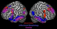
The study found that there are, in fact, distinct regions of the brain that are used to process the two types of sentences: those in which word order determined the relationships between the sentence elements, and those in which inflection was providing the information.
In fact, Newman said, in trying to understand different types of grammar, humans draw on regions of the brain that are designed to accomplish primitive tasks that relate to the type of sentence they are trying to interpret. For instance, a word order sentence draws on parts of the frontal cortex that give humans the ability to put information into sequences, while an inflectional sentence draws on parts of the temporal lobe that specialize in dividing information into its constituent parts, the study demonstrated.
"These results show that people really ought to think of language and the brain in a different way, in terms of how the brain capitalizes on some perhaps preexisting computational structures to interpret language," Newport said.
Aside from providing perspective on how language abilities might have evolved in humans, the scientists' findings could perhaps eventually find applications in medicine, according to Newport. For instance, it could prove valuable in assessing how best to teach language to a person with brain damage in certain areas but not others, such as a stroke victim.

How Exercise Might Help Keep Alzheimer's At Bay

 People have been trying for years to keep their brains sharp by exercising, staying mentally active and watching their diets. But a panel convened by the National Institutes of Health warned the public Wednesday that it's not clear whether any of these measures can prevent Alzheimer's disease or other forms of mental decline in people.
Many scientists are still optimistic about prevention, partly because they are also considering research done on animals.
At about the time the panel was releasing its report, a 78-year-old senator was doing something he hopes is good for his brain.
Sen. Richard Lugar (R-IN) was competing in an annual charity race a few miles from Capitol Hill. He's been a runner since grade school and says he thinks exercise helps him remember a lot of stuff, including "the names and places of thousands of people and events that I bring up frequently in the course of debate.
"It's very helpful to have that kind of historical knowledge of my constituency, as well as of the world," Lugar said.
The panel convened by the NIH wasn't so sure that's why people like Lugar remain sharp into their 70s and 80s. And the panel wanted to make sure the public isn't being misled about the benefits of this or any other strategy for preventing Alzheimer's.
So panel members looked only at studies in humans, and they found that some studies of exercise in people have found a benefit while others haven't.
Arthur Kramer, a neuroscientist from the University of Illinois, wasn't on the panel, though he was invited to speak to the group.
He said the panel is right to be cautious, but that it also makes sense for researchers to talk about the potential of exercise.
"The benefits tend to be on the order of a 20 to 30 percent reduction in being diagnosed with Alzheimer's disease and other such diseases," Kramer said. "And again, this isn't universal but this is found in an increasing number of studies."
Kramer said researchers also tend to consider studies that show what exercise does for animals.
"There are improvements in the chemistry of the brain in terms of the molecules that protect the brain, increases in the number of connections between neurons, which allows us to encode new learning and memory, and even the birth of new neurons in one region of the brain that supports memory," he said.
Mental exercise is another strategy that seems like a good idea to many Alzheimer's researchers. After all, it appears to increase connections in the brain and perhaps make the brain more resilient — in animals and perhaps in people.
But Neil Buckholtz from the National Institute on Aging says the panel would need much more than that to recommend a specific activity to the public.
"Doing crossword puzzles, Sudoku, those kinds of things — they're interesting, but the evidence is not available at this point that they actually have an effect," Buckholtz said.
The panel seemed most skeptical about studies of drugs, diets and nutritional supplements.
Members found some evidence of benefit from omega-3 fatty acids like those in fish. But it found no convincing studies in people that antioxidants like vitamin E could make a difference.
Martha Clare Morris, a nutritional epidemiologist at Rush University Medical School in Chicago, is less skeptical. She says there is good evidence that some antioxidants work in animals.
"There's a very broad base of animal models showing that vitamin E protects the brain from neuron loss, from DNA damage, from oxidative damage," she said.
Panel members say they will consider changing their position on vitamin E and other popular prevention strategies when researchers show they work in people.

Brain cell study sheds light on epilepsy

PHILADELPHIA, April 29 (UPI) -- U.S. neuroscientists have identified a process that helps control the firing of neurons -- a finding they said sheds light on epileptic seizures.

The researchers from the Children's Hospital of Philadelphia and Tufts University School of Medicine in Boston said by isolating the molecular and electrical events that occur when the control is disrupted, the new research not only advances medicine's understand of epileptic seizures, but also potentially other diseases involving poorly regulated brain activity.

"By better understanding the detailed events that occur in epilepsy, we are gaining knowledge that could ultimately lead to better treatments for epilepsy, and possibly for other neurological diseases," said Children's Hospital neuroscientist Douglas Coulter, the study's corresponding author. "Temporal lobe epilepsy, in particular, often resists current treatments," he said.

Coulter's research group, collaborating with a Tufts University team led by co-senior author Professor Philip Haydon, explained that in epilepsy excessive signaling between neurons can lead to seizures. However, another class of brain cells called glia can regulate such signals. Among the glia are star-shaped cells called astrocytes -- the particular focus of this research.

"This study shows that changes in astrocytes are key to brain dysfunction and opens the potential for novel therapeutic strategies in epilepsy," Haydon said.

The study is detailed in the early online edition of the journal Nature Neuroscience.

Docs pull out 8cm splinter from boy's brain

NEW DELHI: The doctors were shocked when 11-year-old Rahul walked into the neurosurgery OPD at AIIMS a few days ago. Rahul had been walking about with an 8cm-long wooden splinter in his brain it had pierced his left eye for the past two years. On Wednesday, doctors at AIIMS extricated, in a five-hour-long surgery, the splinter from Rahul's brain. The doctors were very surprised that Rahul hadn't developed a neurological or other complication over the past two years.

"It's unbelievable. Despite having a foreign body in his brain, he didn't develop a medical complication. He was absolutely fine when he came to us,'' said Dr P Sarat Chandra, associate professor, AIIMS, who operated on Rahul.

According to Rahul's family, he was playing when he fell on a wooden stick, a splinter from which pierced his left eye and entered his brain. "The stick broke when he tried to get up. We took him to a local hospital where doctors said they couldn't remove it,'' said Ratna Devi, Rahul's mother, who took him to several hospitals before bringing him to AIIMS. Because of the accident, Rahul lost his vision in the left eye but that didn't stop him from going to school and going about his everyday activities.

Apart from the left eye orbit, the splinter pierced the temporal lobe (responsible for memory), cavernous sinus (a collection of thin-walled veins at the base of the brain), the cerebellum (responsible for motor function), and was exerting pressure on the brain stem. "The splinter missed the internal carotid artery, which is very close to the cavernous sinus, by less than a millimetre. Since the splinter had been lodged in the brain for two years, there was some inflammation around it and there were significant changes near the cerebellum. Had the splinter been pushed farther inside, it could have been fatal,'' said Chandra.

Doctors say Rahul's case is nothing short of a miracle. "All the major areas in the brain were affected by the splinter, but there were no complications. It is surprising that he didn't develop an infection over the past two years. And he is very lucky there was no major damage to the brain,'' said Dr AK Mahapatra, head of the department of neurosurgery, AIIMS.

During the surgery, doctors had to be very careful while pulling the splinter out. Even a minor error might have proved fatal. "We cut open the front of the left skull and the roof of the eye orbit. We retracted the brain so that we could see the splinter and then, slowly, pulled it out. The splinter had become soggy and it was difficult to pull it out in one piece. We then washed the affected areas of the brain with antibiotic solution,'' said Dr Chandra.

Doctors say that it will take Rahul a month or two to recover completely. "The risk of him catching an infection is high. He can still get meningitis so we have to be very careful,'' said Dr Mahapatra.

First McCullom Lake Brain Cancer Lawsuit Trials to Begin in June

feature photo The first of more than two dozen toxic tort lawsuits filed on behalf of individuals diagnosed with brain cancer after living in the area of McCullom Lake, Illinois are set to go to trial in early June.
The McCullom Lake brain cancer lawsuits have been filed against various defendants, including Rohm and Haas, which operated a chemical plant in the area. While many defendants have settled out of court, the first trial against Rohm & Haas and other non-settling defendants is scheduled to begin in Philadelphia in a little over one month.
About 30 people out of a community of only 1,000 people have been diagnosed with various forms of brain cancer in recent years. The cancer rate in the community is extremely high, considering the national average is only 7 brain cancer cases per 100,000 people.
The number of brain cancer lawsuits for McCullom Lake residents has grown steadily since the first cases were brought in 2006.
Several of the cases were diagnosed through a $1.4 million cancer cluster class action lawsuit settlement reached with nearby Modine Manufacturing, Inc. in 2008. The original settlement fund has been used to provide numerous vouchers for pre-paid medical testing to past and present residents of the area to screen for brain cancer and brain tumors. At least two of the cases were detected through MRI scans performed for residents using the settlement medical vouchers.
Plaintiffs’ attorneys have said, despite the approaching trial, that there still could be more lawsuits filed as residents who have suffered injuries step forward.
The lawsuits contain allegations that Morton International and Rohm and Haas dumped chemicals illegally into local groundwater for decades, causing the unusually high cancer rate. Morton International owned the McCullom Lake chemical plant until 1999, when it was bought by Rohm and Haas. Rohm and Haas was purchased by Dow Chemical in April of last year.
Despite having admitted that chemicals were dumped in an eight-acre unlined pit for 20 years, Rohm and Haas has pledged to fight the lawsuits, saying their tests showed that contamination from the dumping flowed away from the village. In addition, the county tested 14 local wells and found no water contamination. However, plaintiffs contest the company’s claims about the flow of contaminants, and point out that the county tested the wells years after the company had ceased dumping, and never tested those wells while the groundwater was being contaminated.
Residents of another small town in Missouri have experienced a similar cluster of cancer, resulting in a number of brain tumor lawsuits for residents of Cameron, Missouri that allege the problems were caused by a tannery distributing toxic waste sludge to farmers for use as fertilizer on their fields. At least 70 Cameron, Missouri brain tumor cases have been diagnosed in that community of about 10,000 people.

Sex hormones control masculinization

Couple kissing.jpg
Sex hormones controls ‘masculinization’ of the brain
A new study has uncovered some information about how sex hormones control masculinization of the brain during development and drive gender related behaviors in adult males.

Published by Cell Press in the April 29 issue of the journal Neuron , the study demonstrates that direct action of testosterone, the prototypical male hormone, is unnecessary for masculinizing the brain and behavior.

Testosterone and estrogen are thought to play an essential role in organizing and activating gender-specific patterns of behavior in sexually reproducing animals.

Testosterone is produced by the testes and directly activates the androgen receptor (AR) in target tissues such as muscle. Estrogen is produced by the ovaries and is nearly undetectable in the circulation of males of most species. However, circulating testosterone in males can be converted into estrogen in the brain, and this testosterone-derived estrogen has been shown to control many male behaviors.

"It was known that testosterone and estrogen are essential for typical male behaviors in many vertebrate species," explains the study’s senior author, Dr. Nirao M. Shah from the Department of Anatomy at the University of California, San Francisco. "However, how these two hormones interact to control masculinization of the brain and behavior remained to be established."

Dr. Shah and colleagues found that during the neonatal testosterone surge there is very little AR expressed in the developing brain, making it unlikely that testosterone signaling via AR plays a major role in masculinizing neural pathways. Importantly, they went on to show that the male pattern of AR expression in the brain was dependent on testosterone-derived estrogen signaling.

The researchers then used a genetic approach to knock out the AR in the mouse nervous system and observed that these mutants still exhibited male type mating, fighting, and territorial marking behaviours.

However, these mutant males had striking reductions in specific components of these masculine behaviors.

These results show that testosterone signaling via AR does not control masculine differentiation of the brain and behavior but regulates the frequency and extent of male typical behaviors.

"Our findings in conjunction with previous work suggest a model for the control of male pattern behaviors in which estrogen masculinizes the neural circuits for mating, fighting, and territory marking, and testosterone and estrogen signaling generate the male typical levels of these behaviors," concludes Dr. Shah. "It will be interesting in future studies to identify the molecular and circuit level mechanisms that are controlled by these hormones."

Supplements in fish oil do not boost brain power

Rejecting the long held belief that supplements in fish oil are good for kids brain, a new study claimed that the pills do not boost mental ability of children.
Supplements in fish oil do not boost brain power1 300x300 Supplements in fish oil do not boost brain power
For the study, which according to the researchers is the largest of its kind, 450 children aged eight to ten at 18 schools in South Wales were given either omega-3 supplements ‘clever capsules’ or placebos for a period of four months.
The results of a series of tests showed that the fish oil pills did not improve the youngsters’Supplements in fish oil do not boost brain power it did appear that those taking them were more attentive.
Further analysis showed that reading, spelling and handwriting were not improved in those who took omega-3, the Daily Mail reported.
Pointing that supplements might help some youngsters who have trouble concentrating in class, lead researcher Amanda Kirby said: “The primary message always has got to be to start with a good diet.”
Noting that kids are eating more rubbish, Kirby of the University of Wales, said: “If children have a relatively varied diet and don’t seem to have problems, it is probably not going to help them.”
For youngsters with attention deficit hyperactivity disorder or learning difficulties, fish oils are “worth a try,” she said.
She added: “Fatty acids make up 20 per cent of the brain and are going to have an effect in a number of different ways. Some of the studies on cardiovascular disease and Alzheimer’s disease are pretty convincing, but we need more research.”
Earlier studies have credited fats found in abundance in fish such as herring, mackerel, salmon and fresh tuna with health benefits from staving off heart disease, cancer and depression, to warding off Alzheimer’s disease.

Parkinson's eased by brain probe

The invasive implant surgery is not suitable for all patients
A brain “pacemaker” can fight Parkinson's disease, according to The Independent. The newspaper said that combining deep brain stimulation (DBS) implant surgery with standard drug treatment has been found to give greater improvement in motor function and to reduce symptoms more than drug treatment alone.
The research behind this news was a trial involving 366 people with advanced Parkinson’s disease that was not being adequately controlled with medication. It found that after a year, those who had a DBS implant had greater improvements in quality of life than those receiving medical treatment alone. This was particularly due to improvements in mobility, bodily discomfort and the ability to carry out the activities of daily living. However, DBS surgery was not without risks, and about 19% of patients had serious adverse effects, mainly infections.
This trial suggests that combining DBS with medication has some benefits beyond drug therapy alone. Importantly, though, DBS treatment is invasive and will not be appropriate for everyone with Parkinson’s. This means that the potential benefits of DBS would need to be balanced against its risks for each patient.

Where did the story come from?

This research was carried out by Professor Adrian Williams and colleagues from the Queen Elizabeth Hospital in Birmingham and other hospitals and research centres in the UK. The study was funded by the UK Medical Research Council, Parkinson’s UK and the Department of Health. It was published in the peer-reviewed medical journal, The Lancet.
The BBC News website, Daily Mail and The Independent covered this story in an accurate and balanced way. The Daily Mail and BBC News reported that this was a decade-long trial, although the trial recruited participants between 2000 and 2006, so a number of the patients will not yet have been followed for a full ten years. The current results are also only based on follow-up in the year after surgery, with longer-term results awaited. The Independent reported that 5% of people receiving DBS had severe complications, such as infections. However, 19% were reported to have serious surgery-related adverse events in the research paper.

What kind of research was this?

This was a randomised controlled trial (RCT) called PD-SURG, which looked at the effect of deep brain stimulation (DBS) on quality of life in people with advanced Parkinson’s disease. Treatment with DBS involves implanting wire electrodes into the brain. These electrodes are attached to a “pacemaker” device, which regularly sends electrical impulses through the electrodes and into the brain. In most cases, the pacemakers in this trial were implanted into an area of the brain known as the subthalamic nucleus, although other DBS procedures may use alternative sites.
An RCT is the most appropriate way to compare the effects of different treatments. This RCT compared the best medical treatment alone with the same type of medical treatment combined with a DBS implant. This study design would be the best way to tell whether DBS provided any additional benefits over and above standard treatment.

What did the research involve?

The researchers recruited 366 people with Parkinson’s disease that was not adequately controlled with medication alone. They were randomised to continue to receive best medical treatment alone (drugs such as dopamine agonists, MAO type B inhibitors, COMT inhibitors and apomorphine) or to receive DBS surgery in addition to the best medical treatment. The researchers followed the participants up for one year and measured their quality of life to see whether DBS had any effect on this outcome.
The participants in this trial were enrolled at 13 neurosurgery centres in the UK between 2000 and 2006. They had to have Parkinson’s disease diagnosed according to standard criteria, and to be fit enough to undergo surgery. Before being randomised, the participants filled out a standard Parkinson’s disease questionnaire (PDQ-39), which assessed their quality of life. One year after being randomised and receiving their assigned treatment, the participants filled in this questionnaire again.
The researchers then compared changes in quality of life in the group that received DBS and the group that did not. A change of 10 points on the questionnaire score (based on a 39-point scale) was considered to be large enough to be meaningful to patients. A secondary outcome assessed by researchers was clinical assessment of the participants’ functioning using UPDRS scores, a standard scale for measuring Parkinson’s symptoms.
As one group had surgery and the other did not, it was not possible to blind participants to which treatment they received. The researchers also knew what treatments the participants had received as the study did not have sufficient resources to use independent blinded assessors for clinical assessments. People in the standard treatment group (the non-surgery group) could have surgery after one year if their treatment was still not adequately effective.

What were the basic results?

One year after surgery, people who received DBS in addition to best medical treatment showed greater improvement in their quality of life than those who received best medical treatment alone. The DBS group improved by 5 points on the PDQ-39 scale and the medical group by only 0.3 points.
 The quality of life questionnaire assessed different areas of life and showed that people who received DBS had greater improvements in mobility, activities of daily living and bodily discomfort. The difference between the groups was 8.9 points for mobility, 12.4 points for activities of daily living, and 7.5 points for bodily discomfort. Participants who received DBS also showed greater improvements in clinically assessed overall functioning at one year than participants receiving medication alone. Participants who received DBS had reduced their drug dose by about 34% compared with the medical treatment group.
Just under one in five people who received DBS had serious adverse effects associated with their surgery (19%), and one patient died from bleeding during surgery. Similar proportions of patients had side effects of their medical treatment in both groups (11% with DBS plus medical treatment, and 7% with medical treatment alone).

How did the researchers interpret the results?

The researchers concluded that one year after the study began, treatment that combined surgery and best medical therapy “improved patient self-reported quality of life more than best medical therapy alone in patients with advanced Parkinson’s disease”.
They also say that the improvements seen were clinically meaningful, but that the risks associated with DBS surgery may warrant only offering the surgery to those people most likely to benefit from it.

Conclusion

This study used a robust design to assess the effects of deep brain stimulation (DBS) on quality of life in people with Parkinson’s disease that had not responded adequately to medical treatment. Points to note include:
  • Blinding participants and researchers to the treatment received was not possible, so participants’ ratings of their quality of life may have been affected if they had pre-existing expectations of DBS or if they were disappointed not to have received DBS.
  • The trial has so far collected and reported one year’s worth of data. The researchers are continuing to collect information on the patients’ outcomes so that the longer-term effects of DBS can be studied.
  • The researchers suggest that the group of patients treated were representative of those who would be offered surgery at neuroscience centres in the UK.
  • A questionnaire was given to participants in the DBS group about surgery-related adverse effects six months after surgery, but a similar questionnaire was not given to the medical treatment only group. Therefore, adverse effects in the latter group could have been missed. The researchers also note that they did not record adverse effects that were not serious enough to cause a patient to be admitted to hospital or to extend their stay in hospital.
  • People who received DBS continued to receive medical therapy, although the drug dose could be reduced in many cases. Therefore, news reports that “brain surgery is more effective than medication” or “implants have given us our life back” should not be misinterpreted to mean that DBS is a complete cure or that a person will no longer need any form of drug treatment. People should also be aware that all surgical procedures are associated with some degree of risk and this treatment would not be suitable for everyone. Advances and developments in the DBS technique are likely to continue.
Overall, the results suggest that combining DBS with the best medical therapy can improve quality of life more than medical treatment alone in people with Parkinson’s disease that has not responded adequately to medical treatment.