Brain tumour's 'grow-or-go' switch found (Getty
American researchers have discovered the brain tumour switch responsible for the 'grow-or-go' phenomenon.
Cancer cells in brain tumours have to adjust to periods of low energy or die. When energy levels are high, tumour cells grow and multiply but when levels are low, the cells grow less and migrate more.
Scientists at the Ohio State University Comprehensive Cancer Center-Arthur G James Cancer Hospital and Richard J Solove Research Institute discovered that a molecule called miR-451 directs the change, and that the change is accompanied by slower cell proliferation and an increase in cell migration.
This behaviour was closely associated to the cancer's ability to invade and spread. Thus, the molecule might be used as a biomarker to predict how long patients with the brain tumour glioblastoma multiforme will survive and may serve as a target to develop drugs to fight these tumors.
The researchers found that glioblastoma cells shift from their typical means of metabolizing glucose, a sugar brought by the bloodstream and usually used for energy, to an alternate means that consumes resources within the cell.
Co-author Dr E Antonio Chiocca, professor and chair of Neurological Surgery at Ohio State, said: "Our study reveals how brain tumor cells adapt to their surroundings and survive conditions that might fatally starve them of energy.
"We have discovered that glioblastoma cells use miR451 to sense the availability of a nutrient - glucose. Levels of miR-451 directly shut down the engine of the tumor cell if there in no glucose or rev it up if there is lots of glucose. This important insight suggests that this molecule might be useful as a biomarker to predict a glioblastoma patient's prognosis, and that it might be used as a target to develop drugs to fight these tumors."
The tumours are highly invasive, which makes them difficult to remove surgically, and respond poorly to radiation therapy and chemotherapy.
Average survival for patients is 14 months after diagnosis. MiR-451 belongs to a class of molecules called microRNA, which play a key role in regulating the levels of proteins that cells make. Changes in levels of these molecules are a feature of many cancers, according to the researchers.
Principal investigator Sean Lawler, assistant professor of neurological surgery, said: "The change in miR-451 expression enabled the cells to survive periods of stress caused by low glucose, and it causes them to move, perhaps enabling them to find a better glucose supply.
The migration of cancer cells from the primary tumor, either as single cells or as chains of cells, into the surrounding brain is a real problem with these tumors. By targeting miR-451, we might limit the tumor's spread and extend a patient's life."
For the study, Lawler, Chiocca, Jakub Godlewski, the postdoctoral fellow who was the first author of the study, and their team first compared microRNA levels in migrating and nonmigrating human glioblastoma multiforme cells. The analysis suggested an important role for miR-451.
Experiments with living cells demonstrated that high levels of glucose correlated with high levels of the molecule, and that this promotes a high rate of tumor-cell proliferation. Low glucose levels, on the other hand, demonstrated cell proliferation and increased cell migration.
Moreover, when the scientists boosted levels of the molecule in migrating cells, it slowed migration 60 per cent, and, after 72 hours, almost doubled the rate of cell proliferation compared with controls.
Interestingly, when they forced an increase in miR-451 levels, the cells quickly died, suggesting a possible role in therapy.
Analyses of patient tumours demonstrated that three of five had elevated levels of the molecule. Finally, the researchers compared the survival in 16 patients with high miR-451 and 23 patients with low levels. Those with high levels of the molecule had an average survival of about 280 days while those with low levels lived an average of about 480 days.
Chiocca said: "This suggests that molecule may be a useful prognostic marker."
The findings of the study have appeared in the March 12 issue of the journal Molecular Cell.
Saturday, March 13, 2010
Brain Activity Analysis Reveals Memories
They leave a trace in the cortex as they unfold

Experts investigating the way in which our brain forms, stores and recalls memory were recently able to demonstrate through imaging techniques that these events leave a trace in the cortex. The real finding is that this trace can be viewed with existing equipment. The discovery could lead to a better understanding of neurological conditions, as well as to the development of potential new cures. Memory impairments, such as those produced by a stroke, an injury, or simply by aging, could also become a thing of the past, the team behind the study is quoted by ScienceNow as saying.
The investigation was conducted by the same researchers who in a previous study determined the secrets of the hippocampus. This extremely important area of the brain is capable of keeping tabs on where a person is at all times, and is also crucial in learning and memory. Using a proprietary algorithm, as well as the perks of functional Magnetic Resonance Imaging (fMRI), British researchers from the University College London (UCL) managed to crack its secrets. The team was led by renowned international expert and cognitive neuroscientst, Eleanor Maguire.
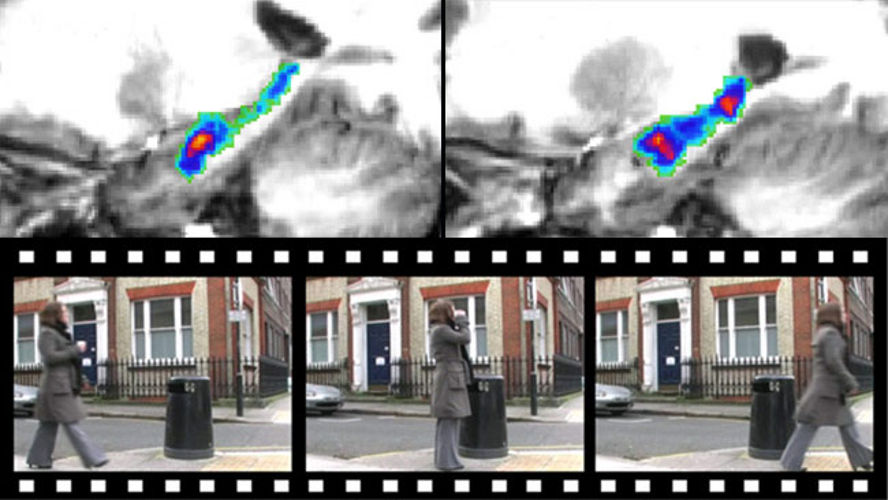
In the new experiments, Maguire and her team shifted their attention from spatial orientation to another, more complex function of the hippocampus. It is called episodic memory of specific experiences, and the group says that a good example for this is the associations formed inside the brain when an individual sees the ocean for the first time. The team wanted to learn whether using their algorithm could allow for the capturing of such memories. The experts selected ten volunteers that shared the memory, and then placed in them in fMRI machine. The imaging method highlights which areas of the brain activate in the presence of a certain stimuli, by analyzing blood flow.
The test participants were shown three different movies, each of them 7 seconds long. They were asked to remember what they saw on a screen while inside the fMRI machine. The computer algorithm was then put to work to determine possible associations between their brain activation patterns. In the end, the computer code managed to identify which of the movies the participants were remembering with an accuracy “considerably higher than would be expected by chance,” Maguire says. “This is really an interesting result, […] it's the closest we've come to reading specific memories,” adds of the work University
Subscribe to:
Comments (Atom)