Thursday, October 18, 2012

Inside the Brain: A Journey Through Time

The Human Brain
The brain has long boggled the mind with its complexity, which is probably best summed up by Carl Sagan in "The Cosmos," when he said, "The brain is a very big place in a very small space." With modern technology, scientists are peering deeper and closer than ever before at the tangle of neurons and their billions of connections. Here's a peek at what the brain looks like, from antiquity to present-day.

Portraits of the Mind
In the book, "Portraits of the Mind: Visualizing the Brain from Antiquity to the 21st Century" (Abrams 2010), astonishing images that reveal both the complexity and beauty of the brain. And through time as brain-imaging technology comes online, scientists have new ways of seeing and interpreting the brain. Check out some of the amazing photos from the book.

Canine Scents
This 1875 drawing showing a dog's olfactory bulb was completed using a staining method named after Camillo Golgi in which certain chemicals are injected into nervous tissue so they can be seen. Some say its application to the study of brain tissue represents the beginning of modern neuroscience.

Dripping Dendrites
While all cells in the body hold the same genome, only a particular set of its genes get turned on in various cells; each type of neuron switches on a gene set that defines its character.
In this picture, a gene called JAM-B had been switched on, which then turned on a fluorescent protein to reveal a small group of brain cells. The resulting image shows that all of the neurons' projections called dendrites are aligned in the same direction; moreover, these retinal neurons are known to detect only objects moving in an upward direction.

Baroque Blood Vessels
A scanning electron microscope (SEM) image zooms in on the baroque branching structures that send blood to the human brain's cortex. The vessels are organized such that the large blood vessels surround the surface of the brain (top of image), sending thin, dense projections down into the depths of the cortex (bottom of image).

View of a StrokeCredit
A brain-imaging method called diffusion MRI (magnetic resonance imaging) is relatively new to the field of neuroscience, though it shows promise as a diagnostic tool. Here, an image taken from the brain of a patient who suffered a stroke in the thalamus and midbrain, resulting in damage to certain axons (some are visible at the bottom of the image).

Mouse BrainCredit
A cross-section of a mouse's hippocampus — one of the brain's memory centers — reveals its intricate network of neurons, whose soma are shown as small circles. The hippocampus is seen here nestled directly beneath the neocortex, which is the outer layer of the cerebral hemispheres.

Spiny NeuronCredit
Most neurons have three parts: an axon, a cell body called a soma and dendrites. This scanning electron microscope (SEM) image shows a soma with dendrites (and their spines) radiating from it. To create SEM images, a beam of electrons is scanned across the surface of a sample, and a detector keeps track of electrons bouncing off its surface to reveal the specimen's outer shape.

Artsy Brain CellsCredit
Here, two types of cells in the cerebellum are shown: glia and Purkinje neurons. The cells can be distinguished because of a method that relies on the body's immune system and its antibodies — proteins that recognize and latch onto "foreign substances." Biologists now use antibodies to reveal where certain proteins are found in the brain. Here, red is an antibody staining of a protein that's found in glia cells, while green reveals a protein called IP3, of which Purkinje neurons are chockfull.

Color My Cerebellum The colored splotches reveal so-called presynaptic terminals, or junctions through which neuron signals are sent, formed by the cerebellum's axons.

BrainbowCredit
While Golgi's staining method did wonders for finding structures hidden in a tangle of neurons, it couldn't distinguish individual brain cells that were illuminated in the same color.
Enter a bit of genetic trickery called Brainbow: Robert Tsien and other chemists tinkered with and discovered fluorescent proteins responsible for the different colors emitted by various sea creatures (such as corals and jellyfish). By coaxing different sets of neurons or even different individuals of a species (say a male and female) to express different proteins, scientists could pick out the cells by the color they glowed.
Here, several motor-neuron axons (slender projections on neurons that transmit signals to other neurons) travel side by side as they lead to the muscles whose contractions they regulate.

Studies Reveal How Diet Affects Brain Functions

TEHRAN (FNA)- Studies released recently explored the neurological component of dietary disorders, uncovering evidence that the brain's biological mechanisms may contribute to significant public health challenges - obesity, diabetes, binge eating, and the allure of the high-calorie meal. 
 
The findings were presented at Neuroscience 2012, the annual meeting of the Society for Neuroscience and the world's largest source of emerging news about brain science and health.

Scientists are ultimately searching for new ways to treat diet-related disorders while raising awareness that diet and obesity affect mental as well as physical health.

Today's new findings show that:

Being obese appears to affect cognitive function, requiring more effort to complete a complex decision-making task (Timothy Verstynen, PhD, abstract 802.20).

Brain images suggest that when people skip breakfast, the pleasure-seeking part of the brain is activated by pictures of high-calorie food. Skipping breakfast also appears to increase food consumption at lunch, possibly casting doubt on the use of fasting as an approach to diet control (Tony Goldstone, MD, PhD, abstract 798.02).

A study in rats suggests they may be able to curb binge-eating behavior with medication used to keep substance abusers clean and sober (Angelo Blasio, PhD, abstract 283.03).

Other recent findings discussed show that:

Amidst growing concern that diet-related metabolic disorders such as diabetes impair brain function, an animal study reports that a high-sugar diet may affect insulin receptors in the brain and dull spatial learning and memory skills. But omega-3 supplements may at least partially offset this effect (Rahul Agrawal, PhD).

Evidence from a rat study suggests that a new compound under development to treat compulsive eating disorders and obesity may be effective at blocking a specific receptor in the brain that triggers food cravings and eating when activated by "food related cues," such as pictures or smells, irrespective of the body's energy needs (Chiara Giuliano, PhD).

"These are fascinating studies because they show the brain is an often overlooked yet significant organ in an array of dietary disorders," said press conference moderator Paul Kenny, PhD, of The Scripps Research Institute in Florida, an expert on addiction and obesity. "Many of these findings have the potential to lead to new interventions that can help reduce the ranks of the obese, helping those who struggle daily with dietary decisions reassert control over what they eat."

This research was supported by national funding agencies such as the National Institutes of Health, as well as private and philanthropic organizations.

Dolphins stay awake for 15 days by sleeping with one half of brain

Dolphins can stay alert and active for 15 days or more by sleeping with one half of their brain at a time, scientists have learned.

Dolohins can stay away for up to 15 days by sleeping with one half of their brain at a time
Dolphins can stay alert and active for 15 days or more by sleeping with one half of their brain at a time, scientists have learned.
The trick of keeping half the brain continuously awake is vital to the sea mammals' survival, experts believe.
It allows them to come to the surface every so often to breath, and remain constantly vigilant for sharks.
Scientists in California, US, tested the ability of two bottlenose dolphins to echolocate accurately over periods of time which would have left other animals sleep-deprived and exhausted.
The dolphins, a male called Nay and female called Say, had to swim around a pen looking for phantom sonar targets.

Each of the eight targets consisted of a device that picked up dolphin sound pulses and sent back ''phantom'' echoes.

When a dolphin detected an echo from an activated target, it responded by pressing a paddle. Correct detection triggered a tone, signalling success, and the dolphin was rewarded with a fish. False alarms led to no tone and no reward.

Over three sessions of five continuous days both dolphins did well, with success rates of up to 99%, but Say outperformed her male partner.

The scientists then went on to test Say further by repeating the same experiment over a period of 30 days. In the event, a storm cut the trial short after 15 days had elapsed. However, during this time Say's performance hardly deteriorated at all.

The findings were published yesterday in the online journal Public Library of Science ONE.

Lead researcher Dr Brian Branstetter, from the National Marine Mammal Foundation in San Diego, said: ''These majestic beasts are true unwavering sentinels of the sea. The demands of ocean life on air breathing dolphins have led to incredible capabilities, one of which is the ability to continuously, perhaps indefinitely, maintain vigilant behaviour through echolocation.''

The difference in performance between Nay and Say was probably down to personality, said the scientists.
Say appeared much more motivated and keen on the task, often producing ''victory squeals'' when she correctly responded to a target.

Dolphins use their sonar-like ability to navigate, find prey, detect predators, and co-ordinate group behaviour.

Brain wave measurements have confirmed that the creatures are capable of ''unihemispheric sleep'' - sleeping with just one side of the brain. When dolphins sleep in this way, they often keep one eye open.
Many dolphin populations are exposed to almost constant risk of shark attack, and the animals commonly carry bite marks, said the researchers. Continually listening to their echo signals allowed them to counter the shark threat, even in murky waters.

The scientists wrote: ''From an anthropomorphic viewpoint, the ability of the dolphin to continuously monitor its environment for days without interruption seems extreme.

''However, the biological, sensory and cognitive ecology of these animals is relatively unique and demanding. If dolphins sleep like terrestrial animals, they might drown. If dolphins fail to maintain vigilance, they become susceptible to predation. As a result, the apparent 'extreme' capabilities these animals possess are likely to be quite normal, unspectacular, and necessary for survival from the dolphin's perspective.''

Scientists discover how brain erases unwanted memories

Scientists have discovered that brain can help us voluntarily forget unwanted memories by either blocking them out or substituting them.

Researchers from the Cambridge University tested if suppressing memories or substituting them with more desirable memories could erase them and whether these tactics could engage distinct neural pathways.

To test this possibility, researchers used functional magnetic resonance imaging to examine the brain activity of volunteers who had learned associations between pairs of words and subsequently attempted to forget these memories by either blocking them out or recalling substitute memories.
Although the strategies were equally effective, they activated distinct neural circuits. During memory suppression, a brain structure called dorsolateral prefrontal cortex inhibited activity in the hippocampus, a region critical for recalling past events. On the other hand, memory substitution was supported by caudal prefrontal cortex and midventrolateral prefrontal cortex - two regions involved in bringing specific memories into awareness in the presence of distracting memories.
"This study is the first demonstration of two distinct mechanisms that cause such forgetting: one by shutting down the remembering system, and the other by facilitating the remembering system to occupy awareness with a substitute memory," said lead study author Roland Benoit.
"A better understanding of these mechanisms and how they break down may ultimately help understanding disorders that are characterised by a deficient regulation of memories, such as posttraumatic stress disorder," Benoit said in a statement.
"Knowing that distinct processes contribute to forgetting may be helpful, because people may naturally be better at one approach or the other," he said.

Training the brain to stress less

Neurotopia recently began beta-testing a dry sensor, mobile headphone and tablet system that would map brain waves.Neurotopia recently began beta-testing a dry sensor, mobile headphone and tablet system that would map brain waves

Editor's note: CNN contributor Amanda Enayati ponders the theme of seeking serenity: the quest for well-being and life balance in stressful times. She delivered a version of this piece as a talk at Stanford's Medicine X conference last week.
(CNN) -- Train the brain. Until recently, this phrase made me picture Neo from "The Matrix" proclaiming "I know kung fu" after he had martial arts abilities uploaded into his brain.

But what if we really could harness technology, Neo-style, to help train our brains to better cope with everyday stress?
For many of us, the days seem to pass in one anxiety-ridden blur after another. Mental health professional increasingly agree that these daily sprints, accompanied by a soundtrack of endless beeps, chirps and vibrations emitting from various devices, set off our stress systems, keeping us in a persistent and physiologically damaging state of fight-or-flight.
"The way we live our lives now is like running marathons," said Dr. Leslie Sherlin, a neuroscientist and chief science officer of Neurotopia, a company that provides brain training to athletes. "And in some ways, that's great, but you can't run marathons all the time."
Keep that pace, says Sherlin, and at some point, you will burn out. You may also suffer from a weakened immune system that can lead to an increased risk of disease.
Most of us have received some kind of formal instruction about diet, exercise, the birds and the bees. So why aren't we training our brains to better manage stress?
Some of the most compelling training to help prepare people to better handle stress is going on right now with athletes and soldiers.
For these two distinct groups, performance under high stress is a must (albeit for very different reasons). But the technologies being used to train them could benefit the rest of us as well.
Technology could help us reduce stress, too
Training athletes for the field
I became interested in the way athletes train for peak performance in high-stakes environments last year, when I interviewed Michael Gervais, a sports psychologist who works with Sherlin to train elite athletes to perform optimally during high-stress competition. Gervais and Sherlin work with athletes from the NFL, NBA and NHL as well as Olympians, golfers and many others.
What Gervais told me then was that the key to high performance was a disciplined mind. While not exactly news, the methods Gervais and his colleagues use to teach mental discipline were quite interesting. They were using older Eastern disciplines like mindfulness, presence, meditation, deep breathing and neurofeedback.
The way we live our lives now is like running marathons. ... In some ways, that's great, but you can't run marathons all the time.   Dr. Leslie Sherlin, chief science officer of Neurotopia
As part of their training, Gervais and his colleagues hook up athletes to electrodes and perform a baseline qEEG: a quantitative electroencephalogram. They use the results to create an individualized brain map.
The map helps these sports psychologists assess and quantify mental aspects of performance like focus, decision speed, reaction time and stress regulation.
Once the brain is mapped, the psychologists conduct half-hour neurofeedback sessions to teach athletes how to reach optimal brain wave patterns. In a typical session, the athlete will sit before a large screen as sensors monitoring electrical activity in his or her brain are placed on the scalp.
The athlete then focuses on achieving desirable brain wave patterns that, in turn, influence what happens on the screen. It's bit like controlling a video game with only your thoughts. The version I saw involved cars racing through a desert.
The training is meant to teach athletes how to respond quickly to stressor stimuli, how to focus during stressful situations, how to recover from errors and finally how to shut down and still their minds when it's all over.
These sports psychologists have collected a proprietary brain bank of assessments over years of working with elite athletes. They use the brain bank to identify optimal brainwave patterns associated with the highest levels of performance.
According to Sherlin, it takes roughly 15 to 20 neurofeedback sessions for elite athletes to learn some of these techniques. (Probably about 30 for you and me, he says.)
Your questions about stress, answered! 
Originally developed as a technique to measure brain activity in NASA pilots during flight simulation exercises, neurofeedback has shown promising initial results for helping retrain the brainwaves of children with ADHD and autism and people suffering from chronic migraines. In one study, student eye surgeons were trained to significantly improve their surgical skills by regulating their own brainwave activity.
The method is being examined in a diverse number of other contexts, including to help relieve symptoms of chemotherapy-induced nerve damage. Controlled, randomized trials will help validate these promising starts.
The kind of training that the athletes working with Gervais and Sherlin receive is not available to most of us right now, but it may be in our near future.
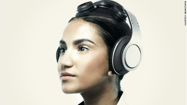
A few weeks ago, Sherlin's company, Neurotopia, began beta-testing a dry (no goo in your hair) sensor, mobile headphone and tablet system that purports to do the same kind of assessment and training as the older model. At least in theory, this might make the product accessible to the rest of us.
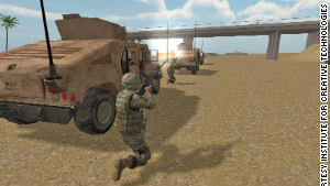
Training soldiers for the battlefield
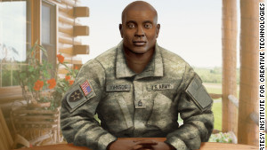
A conversation with Dr. Albert "Skip" Rizzo, psychologist and research professor at the University of Southern California Keck School of Medicine, is like a lesson in applied science fiction, with your mind reeling from "Star Trek" to the original "Total Recall."
Except Rizzo's jaw-dropping efforts are not fiction, nor are they "on the horizon." They are here, now.
In a collaboration between the military, Hollywood and USC's Institute for Creative Technologies, where he serves as the associate director for medical virtual reality, Rizzo and his colleagues have developed cutting-edge gaming and virtual reality technologies to serve the clinical needs of soldiers.
Virtual Iraq (and Afghanistan) are based on exposure therapy, which has been effective in the treatment of PTSD.
One project, Stress Resilience in Virtual Environments (STRIVE), helps train service members to have better resilience and emotional coping skills in realistic virtual-reality combat scenarios before they are exposed to the real stresses of combat.
A second project, called Virtual Iraq (there is also a Virtual Afghanistan), helps soldiers returning from combat work through their trauma by donning a helmet geared with video goggles, earphones and a scent machine, and revisiting the scene in a virtual reality setting, complete with sound and smell. Both STRIVE and Virtual Iraq (and Afghanistan) are based on exposure therapy, which has been effective in the treatment of post-traumatic stress disorder.
The problem with PTSD is that the person often avoids anything that reminds them of the trauma, and this avoidance begins to generalize to everyday things, says Rizzo.
"It's a snowball cascade effect. The things that evoke the fear and anxiety are no longer directly tied to the original trauma but generalized to the outside world. You see people with PTSD who will no longer leave their house, and if they do, they're a nervous wreck."
The idea, says Rizzo, is to re-create the stressful environment in a doctor's office, to help the patients confront and challenge the trauma and to give them the tools to better cope emotionally with what happened.
Both of these technologies require specialists and a clinical setting, but SimCoach, a "virtual human" designed for interactive use on the Internet, does not.
Though at this point, SimCoach is targeted toward active-duty military personnel, veterans and their families, it may also have wider utility for everyday stress and anxiety.
SimCoach users can select one of several avatars to talk to when they are feeling stressed out. The virtual human coaches can serve as an "online companion for anyone who may be too introverted to seek help, someone who may not want to reach out to a clinician or who may feel stigma about seeing a therapist," said John Hart, program manager at the Institute for Creative Technologies.
SimCoach users can select one of several avatars to talk to when they are feeling stressed out
"SimCoach is not a doc-in-the-box, and it's not going to make a diagnosis," Hart observed. Nor is it meant to replace human interaction.
What SimCoach does do is help those suffering from stress and anxiety symptoms begin the conversation about what they may be going through. It may also provide users with more information about what they may be experiencing, suggest local facilities where they can go for care and perhaps even walk them through breathing exercises or stress reduction techniques.
Hart summed up what I find most compelling about SimCoach: "Here we are, sitting on a mountain of valuable information about what to do when you're stressed or feeling depressed. You can see how SimCoach can help people access the right information when they need it."
Imagine the possibilities! An interactive virtual-reality source for information on stress, anxiety and PTSD -- the precursor, perhaps, to a real-life version of "Star Trek's" Emergency Medical Hologram Doctor.
Home sweet home
I recently attended a conference in Portugal. As I made my way through customs at Philadelphia International, a customs agent asked me what I did for a living.
"I write," I said, "mostly about stress."
He stared me down for few moments before saying in a low, gruff tone: "If you really want to understand stress, then you need to spend a day with us here."
And here's the thing: Regardless of what we do, most of us are feeling that same way about our runaway lives. The genie is out of the bottle, and there is little likelihood of us ever going back to a simpler time (if there ever was such a thing).
So, yes, let's discuss technology addiction, always being "on," tech fasting and the need to design devices and apps for greater serenity. But let's also consider how to harness some of these technologies to help us move easier in this new world, Neo-style.

When you're at rest, your brain's right side hums

Brain 2

There's plenty of brain activity even when people are thinking nothing at all. But it's the brain's right side for most people the less-dominant half that stays busiest while you're at rest, according to surprising new findings.

Researchers found that during periods of wakeful rest, the right hemisphere of the brain chatters more to itself than the left hemisphere does. It also sends more messages to the left hemisphere than vice versa. Surprisingly, this remains true whether the owner of the brain is left- or right-handed. That seems odd, because in right-handed people the left hemisphere is the dominant one, and in left-handed people the right is usually more dominant.

Andrei Medvedev of Georgetown University Medical Center's Center for Functional and Molecular Imaging asked 15 study participants to sit peacefully and let their minds drift while they wore a cap that measured brain activity.

This resting state, previously found to improve memory, "is a special state when the brain tries to deal with information that was acquired during previous active states," Medvedev told LiveScience.

The cap, which was covered with optic fibers, shone infrared light into the scalp. The light waves reached the outermost brain layers and bounced back. The amount of light that reflected back told researchers how muchoxygenated and deoxygenated blood was in a specific brain region. Changes in blood oxygenation tell the researchers which brain areas are using more oxygen and are thus more active.

Medvedev was interested in communication within and between brain regions. He found that the right hemisphere was interacting more with itself and with its counterpart than the left hemisphere was.

"I did not expect that," he said. "I actually was expecting that the left hemisphere would be more important, more integrated, but it appears the right hemisphere during this resting state is more connected." [7 Reasons to Meditate]

So far Medvedev and his colleagues don't know why the right hemisphere is so busy. Because the brain goes into a sort of "housecleaning" mode during resting states, it's possible the right hemisphere works as something like an outside housekeeper, organizing and integrating as well as sending that information to the usually dominant left hemisphere, he said.

The research highlights the need to get more lefties into scientific studies, Medvedev said.

"Most brain theories emphasize the dominance of the left hemisphere, especially in right-handed individuals, and that describes the population of participants in these studies. Our study suggests that looking at only the left hemisphere prevents us from a truer understanding of brain function," he said in a statement.

Medvedev reported his findings Oct. 17 in New Orleans at the annual meeting of the Society for Neuroscience.

Anti-depressants linked to brain bleeding


USA: People using a common class of antidepressants may have slightly increased odds of suffering bleeding in the brain – though the risk is still very small, according to a Canadian study looking at more than 500,000 people.

The antidepressants are known as selective serotonin reuptake inhibitors (SSRIs) and include widely used drugs like fluoxetine (Prozac), sertraline (Zoloft), citalopram (Celexa) and paroxetine (Paxil.)

The SSRIs have been linked to a risk of stomach bleeding, but studies have come to conflicting findings on whether SSRI users have any higher risk of hemorrhagic strokes, which happen when there is bleeding in or around the brain.

For the study, which appeared in the journal Neurology, researchers pooled the findings from 16 past studies involving more than 500,000 people who were on SSRIs or not.

Overall, antidepressant users were about 40 to 50 per cent more likely to suffer bleeding in or around the brain.

But while those numbers might sound big, the risks to any one person would be extremely low, said lead researcher Daniel Hackam, an associate professor of medicine at Western University in London, Ontario, Canada.

Based on these figures, he said, there would be one brain hemorrhage for every 10,000 people using an SSRI over one year.

What’s more, the findings do not prove that the antidepressants directly cause brain bleeds. It’s possible, Hackham said, that SSRI users are sicker than non-users or have habits that put them at greater stroke risk.
The researchers tried to account for those factors in their calculations, but some of the studies they analyzed lacked key information, such as peoples’s smoking and drinking habits, and whether they had diabetes.
We can’t infer cause and effect from this, Hackam said.

On the other hand, there are reasons to believe it’s the medications themselves. For one, the hemorrhage risk seemed greatest in the first months after people started using an SSRI.

There’s also a biological argument. SSRIs seem to make it harder for blood cells called platelets to clump together and form clots – and there can be a big drop in a person’s platelet functions in the first weeks after starting an SSRI, he said.

Still, he stressed that people on the antidepressants should not be alarmed.

I think that overall, these medications are quite safe, he added.

But people who are already at increased risk of a brain hemorrhage may need to be careful. That includes people who have had a brain bleed in the past, or are on medications that reduce blood clotting.