Saturday, November 10, 2012

ADHD Drugs Impact The Brain's Reward System

http://www.scientificamerican.com/media/inline/do-adhd-drugs-take-a-toll_1.jpg
Two to three percent of children in denmark meet the standards to be diagnosed with ADHD, making it extremely important to understand how ADHD drugs work. Now, University of Copenhagen researchers are gathering new information about the impact of ADHD medicine by utilizing a new mathematical reconstruction of a small part of a particular brain region which processes reward and punishment, which always involves the chemical dopamine.

Jakob Kisbye Dreyer, postdoctoral candidate at the Department of Neuroscience and Pharmacology, Faculty of Medical and Health Sciences, University of Copenhagen, said:
"It had been discussed for years whether treating ADHD with Ritalin and similar drugs affects the reward system to any significant degree, simply because the dosage given to patients is so low. We are the first to show that some components of the dopamine signaling pathways are extremely sensitive to drugs like Ritalin. We have also developed a unified theory to describe the effect of such drugs on the dopamine signal."
Dreyer stresses in the new study, which was published in the Journal of Neurophysiology, the significance of understanding what happens during treatments with ADHD medications, such as Ritalin, because knowledge helps to develop more advanced drugs, and also to comprehend the psychology behind ADHD.

Human behavior is driven by unconscious assessment of the cost to gain ratio. The new findings demonstrate that ADHD drugs lessen the signals regarding expected consequence or punishment.

Dopamine, a chemical found in the brain, assists in several processes which alter human behaviors. Certain activities, such as having sex, taking narcotics, winning a competition, and eating, boost levels of dopamine being released. The researchers believe that dopamine plays a part in urging us to repeat behaviors that had, in the past, been linked to reward.

Dreyer explained:
"Control mechanisms in the brain help keep the dopamine signal in balance so we can register the tiny deviations that signal reward and punishment. We discovered while trying to describe these control mechanisms that our model can be used to examine the influence of Ritalin, for example, on the signal. Suddenly we could see that different pathways of the reward system are affected to different degrees by the medicine, and we could calculate at what dosage different parts of the signal would be changed or destroyed."
Ritalin and other ADHD medications have been seen to have inconsistent results, because high dosage increases activity and low dosage decreases it, often making it difficult to find the right dosage for each individual patient.

"We can explain this double effect using our theory. The dopamine signal in the part of the brain that controls our motor behavior is only affected at a higher dose that the dose usually prescribed for treatment. Also, our model shows that the threshold between a clinically effective dose and too high a dose is very low. That may explain why the small individual differences between patients have a big impact on treatment," concluded Dreyer.

Ties between brain, better buildings examined

Can the buildings we inhabit make us well sooner? Can they make prisoners calmer? Can they impact an autistic child?
http://www.therapytoronto.ca/images/blogpics/BrainMusic.jpg
Scientists and architects will come together today at Scottsdale's Taliesin West to explore the connection between the brain and buildings.

The "Minding Design: Neuroscience, Design Education, and the Imagination" conference is sold out at 250 participants.

Taliesin West officials see the conference as an opportunity to broaden its reach by connecting experts from around the nation. 

Sean Malone, chief executive officer of the Frank Lloyd Wright Foundation, said the scholarly conference embodies the foundation's vision of transforming people's lives through Wright's principles.

 "We believe we have an opportunity and responsibility to help shape architecture and design at the highest level," Malone said, adding that Taliesin West, Wright's winter home and studio campus that now offers tours and houses the Frank Lloyd Wright School of Architecture, is the ideal place to host. "There was nobody more innovative and engaged than Frank Lloyd Wright." 

Cathedrals, theaters and museums are believed to inspire and heighten creativity, but this conference will delve into what is happening in the brain when we're inside buildings and how architects can better serve certain populations and humanity. 

The symposium brings in neuroscientists and architects from around the country to discuss how environment shapes the human experience through brain research and theories while impacting imagination, health and other areas. 

Tricia Anderson, an interior designer in South Bend, Ind., spotted the conference on Taliesin West's Facebook page and decided it was the perfect complement to her pursuit of advanced degrees in psychology and interior design.

"I've always felt that a person's environment not only reflects your personality, but it also has a strong effect on your creativity and inspiration and your overall well-being," she said.

Michael Arbib, director of the University of Southern California Brain Project, charted ways in which designing a building can use knowledge of brain function. He is a speaker at the symposium.

Also a member of the Academy of Neuroscience for Architecture (anfarch.org), Arbib said that it may be years before designing a building with a knowledge of brain function becomes widely practiced. But the connection between the two could result in more fruitful lives.

Studies of Alzheimer's patients may help influence the design of buildings, which could make it easier for them to find their way around. Hospitals may acquire lighting that does not disrupt the body's circadian rhythms at night.

"I'll be making every effort to make it clear to the architects why they should care about the brain and how it relates to design," he said.

Gene mutation behind brain defects identifiedl

Gene mutation behind brain defects identifiedWashington: Mutations in a single gene – that causes intellectual disability and increases the risk of developing autism spectrum disorder - severely disrupts the organization of developing brain circuits during early childhood, a new study has revealed.

This study by scientists from the Florida campus of The Scripps Research Institute helps explain how genetic mutations can cause profound cognitive and behavioural problems.

“In this study, we did something no one else had done before,” Gavin Rumbaugh, a TSRI associate professor who led the new research, said.

“Using an animal model, we looked at a mutation known to cause intellectual disability and showed for the first time a causative link between abnormal synapse maturation during brain development and life-long cognitive disruptions commonly seen in adults with a neurodevelopmental disorder,” he said.

The study focused on a critical synaptic protein known as SynGAP1.

Mutations in the gene that encodes this protein cause disabilities in an estimated one million people worldwide, according to the paper.

“You might think this accelerated development of brain circuits would make you smarter,” Rumbaugh said.

“But the increased excitability actually disorganizes brain development. We think that early maturation of these excitatory synapses disrupts the timing of later developmental milestones. It rains down chaos on this complex process, preventing normal intellectual and behavioural development,” he said.

Brain Has Distinct Activity Pattern When Losing Consciousness During Anesthesia

A new study from the US reveals for the first time, that the brain has a distinct pattern of electrical activity as patients lose consciousness during anesthesia. The pattern shows very slow oscillations, reflecting a breakdown of communication between the different regions of the brain, each of which shows shorts bursts of activity alternating with longer silences.


http://media.tumblr.com/tumblr_md2cdf4GMa1r70663.jpg
The researchers write about their findings in a paper published online first on 5 November in the Proceedings of the National Academy of Sciences.

They hope that by improving understanding of what happens in the brain as it loses consciousness, the study will help anesthesiologists better maintain the right balance between too little and too much anesthetic.

Senior author Patrick Purdon, an instructor of anesthesia at Massachusetts General Hospital (MGH) and Harvard Medical School, says in a statement, clinicians will now know what to look for on the electroencephalograph (EEG) when putting a patient under anesthesia:

"We now finally have an objective physiological signal for measuring when someone's unconscious under anesthesia."
EEG Patterns in Epileptic Patients An EEG is a machine that records electrical activity of the brain through electrodes on the scalp. It measures changes in voltage resulting from the various currents flowing between neurons or brain cells.

For their study, Purdon and colleagues studied epileptic patients who had electrodes implanted in their brains to monitor seizures and were having an operation to remove them.

The patients received a common anesthetic known as propofol and had their brain activity monitored by EEG.

Propofol activates receptors on neurons, in a way that makes the brain cells less active, although exactly how this happens is not clear.

The researchers noticed the EEG showed a distinct pattern at the point where consciousness was lost. This was about 40 seconds after receiving the anesthetic, and was defined by the moment when patients stopped responding to sounds played to them every four seconds.

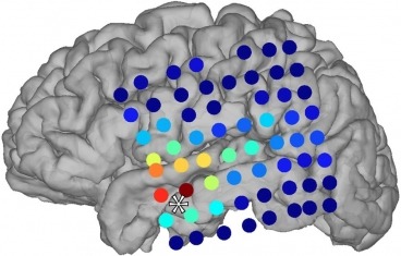
Distinct Pattern of Overall and Local Brain Activity
To record brain activity, Purdon and colleagues used two different sized of electrode, each size taking a different reading of brain activity. The larger electrodes, about the size of a large coin, were placed about 1 cm apart and recorded the overall EEG or brain wave pattern.

The smaller, more localized, electrodes were concentrated in a group of rows about 4 mm wide. Between 50 and 100 of these were implanted in each patient, in different brain regions.

These smaller electrodes recorded activity from individual neurons, and this study is thought to be the first to record neuron activity in patients as they lose consciousness.

The large electrodes showed that within one or two seconds of patients losing consciousness, the EEG pattern suddenly turned to low frequency oscillations, at about 1 cycle per second (about 1 Hz).

This coincided with the small electrodes showing a "flickering" pattern at individual neuron level. Individual neurons within localized brain regions were active for a few hundred milliseconds, then became quiet for a few hundred milliseconds. This created the oscillating pattern seen on the EEG, say the researchers.

"We show that propofol-induced unconsciousness occurs within seconds of the abrupt onset of a slow (< 1 Hz) oscillation in the local field potential. This oscillation marks a state in which cortical neurons maintain local patterns of network activity, but this activity is fragmented across both time and space," they write.

Periodic Silencing Prevents Communication in Brain One of the lead authors, Laura Lewis, a graduate student in the Department of Brain and Cognitive Sciences (BCS) at Massachusetts Institute of Technology (MIT), says:

"Within a small area, things can look pretty normal, but because of this periodic silencing, everything gets interrupted every few hundred milliseconds, and that prevents any communication."

"When one area was active, it was likely that another brain area that it was trying to communicate with was not active. Even when the neurons were on, they still couldn't send information to other brain regions," she explains.
Loss of Consciousness Could Be "Failure of Information Integration"
Michael Avidan is a professor of anesthesiology at Washington University School of Medicine, and was not involved in the study. He describes the findings as "exciting" and suggests they offer neurobiological evidence for the "information integration theory" of consciousness. This theory suggests large-scale brain networks integrate information from the senses to generate our overall impression of the world around us.

When we lose consciousness, there could still be information "coming into the brain, but that information is remaining localized and doesn't get integrated into a coherent picture," he explains.

Another lead author, Emery Brown, professor of brain and cognitive sciences and health sciences and technology at MIT and an anesthesiologist at MGH, says this mechanism of "failure of information integration" has been put forward before as a possible explanation for loss of consciousness, but it was not clear how it worked.

"This finding really narrows it down quite a bit. It really does, in a very fundamental way, constrain the possibilities of what the mechanisms could be," he adds.

Successful Anesthesia: Maintaing a Delicate Balance
The researchers hope the pattern will help anesthesiologists improve monitoring of patients as they receive anesthesia, thus preventing rare cases where patients wake up during operations or where too much anesthetic stops them breathing.

At present, anesthesiologists monitor patients under anesthetic with recordings that calculate an index from the EEG. But that index can hide the underlying physiology that can be seen directly in the slow waves.

Brown says their findings suggest they should be looking at and interpreting the oscillations in the raw EEG readings.

"If you do that, you have a physiologically linked way to know when someone is unconscious. We can take this into the operating room today and give better patient care," he adds.

The team is now going to look at what happens in the brain as it regains consciousness. They have already started looking at the effects of other anesthesia drugs, to see if they generate the same brain patterns.

Purdon says based on EEG studies there appear to be many other drugs producing the same slow oscillations. But there are also a number that are "doing something totally different," he adds.

Funds from the Nationa Institutes of Health (NIH), the Canadian Research Foundation, and the National Institute of Neurological Disorders and Stroke, helped finance the study.

How brain processes information

Washington, November 10 (ANI): A new pathway that seems to play a major role in information processing in the brain has been identified by scientists at the Scripps Research Institute (TSRI).
http://t1.gstatic.com/images?q=tbn:ANd9GcRdfDkYpMp8M9oLtoFLJPcKALFkct6Gz50DqFd83Q7NkzIgcro0f_Q21ijGLw
Their research also shed light on how imbalances in this pathway could contribute to cognitive abnormalities in humans.

The study focuses on the actions of a protein called HDAC4. The researchers found that HDAC4 is critically involved in regulating genes essential for communication between neurons.

"We found that HDAC4 represses these genes, and its function in a given neuron is controlled by activity of other neurons forming a circuit," said TSRI Assistant Professor Anton Maximov, senior investigator for the study.

Richard Sando III, a graduate student at the TSRI Kellogg School of Science and Technology, a member of the Maximov lab and the first author of this study, noted the team become interested in class IIa histone deacetylases (HDACs), which include HDAC4, in part because they have been implicated in regulation of transcription of non-neuronal tissues.

"Class IIa HDACs are also known to change their cellular localization in response to various signals," he said.

"There were hints that, in neurons, the translocation of HDAC4 from the nucleus to cytoplasm may be triggered by synaptic activity. We found that mutant mice lacking excitatory transmitter release in the brain accumulate HDAC4 in neuronal nuclei. But what was really exciting was our discovery that nuclear HDAC4 represses a pool of genes involved in synaptic communication and memory formation," he stated.

Coincidentally, Maximov had been familiar with these same genes since his postdoctoral training with Tomas Sudhof, a neuroscientist whose pioneering work resulted in the identification of key elements of the transmitter release machinery.

"It was truly astonishing when their names came up in our in vitro genome-wide mRNA profiling screens for neuronal HDAC4 targets," Maximov said.

To learn more about the function of HDAC4 in the brain, the team wanted to study its role in a mouse model. First, however, the scientists had to overcome a serious technical obstacle-HDAC4 also appears to protect neurons from apoptosis (programmed cell death), so complete inactivation of this gene would lead to neurodegeneration. To solve this problem, the team generated mice carrying a mutant form of HDAC4 that could not be exported from the cell nucleus. This mutant repressed transcription independently of neuronal activity.

Another surprise came after the team had already initiated their experiments. Underscoring the team's findings, a human genetic study was published linking mutations in the human HDAC4 locus with a rare form of mental retardation.

"One of these human mutations produces a protein similar to a mutant that we introduced into the mouse brain. Furthermore, our studies revealed that these mice do not learn and remember as well as normal mice, and their memory loss is associated with deficits in synaptic transmission. The pieces came together," said Maximov.

The findings have been published in the latest issue of the journal Cell.

Brain tumour survivor in line for a national award

A BRIAN tumour survivor is in line for a national award.

Figure skater Lisa Armitage, of Grimsby, has been shortlisted in the Lifetime Learner Achievement Awards.

Finalist:  Lisa Armitage is one of 20 finalists shortlisted for the Lifetime Learner Achievement Awards.Finalist: Lisa Armitage is one of 20 finalists shortlisted for the Lifetime Learner Achievement Awards.
The 31-year-old – who, as reported, had a tumour the size of a tennis ball removed from her brain last year – will find out if she has won during a glamorous dinner at the Hilton Hotel, Coventry, on Thursday.

The award recognises and celebrates the learning achievements, resulting career and personal successes of those who have completed a qualification with national training provider, Lifetime.

Lisa, who was one of 167 people nominated for the award from across the country, said that fighting the illness made her realise she wanted to get more out of life.

She is now one of 20 finalists in line for the award.

She said: "I feel honoured to have been shortlisted for this award.

"I wanted to try and lead as normal a life as possible through treatment so I completed some training through Lifetime.

"I gained my NVQ level 3 in business and administration while fighting my brain tumour.

"I am currently working as the finance manager at Grimsby Leisure Centre and also doing my NVQ level 3 in leadership and management. The training gave me something else to focus on other than my illness.

"I believe it helped with my recovery."