Friday, March 12, 2010

First Step in Mind Reading Via Computer Program

Machines that decode your thoughts aren't limited to the realm of science fiction anymore.

A computer program that analyzes brain scans was able to tell which of three short films people were thinking about, according to a study in the journal Current Biology.

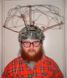
(Jon Wilson has been testing his mind-reading device since the 1980's.)
 
"We were able to predict just from their brain activity which of those memories they were recalling," says Eleanor A. Maguire, one of the study's authors and a professor of cognitive neuroscience at University College London.

This is a major step forward, Maguire says. But it falls short of what most people would call mind reading. "We can't put somebody in a brain scanner and immediately know what thoughts they are having," she says.

Unlocking Our Memories

The experiment was designed to learn more about a part of the brain 
called the hippocampus, which seems to act as a sort of index for memories of events in our lives, Maguire says.

Library of CongressWhen this poster was printed in 1900, mind reading was still in the realm of magic. A new computer program capable of predicting individuals recollections has brought telepathy a small step closer to science.

Machines that decode your thoughts aren't limited to the realm of science fiction anymore.
A computer program that analyzes brain scans was able to tell which of three short films people were thinking about, according to a study in the journal Current Biology.
"We were able to predict just from their brain activity which of those memories they were recalling," says Eleanor A. Maguire, one of the study's authors and a professor of cognitive neuroscience at University College London.
This is a major step forward, Maguire says. But it falls short of what most people would call mind reading. "We can't put somebody in a brain scanner and immediately know what thoughts they are having," she says.
Unlocking Our Memories
The experiment was designed to learn more about a part of the brain called the hippocampus, which seems to act as a sort of index for memories of events in our lives, Maguire says.
She and her colleagues wanted to know whether traces of these so-called episodic memories could be detected in brain scans.
So they found 10 volunteers who agreed to watch several very short films. One showed a woman taking a letter out of her handbag and putting it in a mailbox. In another clip, a woman finishes a cup of coffee and throws the cup in a trash can.
The volunteers watched the films over and over, until they had formed a strong memory of each episode. "Then we popped the people in the scanner and had them recall these movies," Maguire says.
Data from these scans of activity in the hippocampus was analyzed by a computer program that looked for distinct patterns of activity associated with each film. And Maguire says the program found them. "In every single case it was able to predict with high accuracy which of the memories those 10 participants were recalling," she says
That was what the scientists expected to find. What surprised them, though, was how similar the patterns were across all 10 brains. The brain activity associated with each film clip "was incredibly consistent," Maguire says.
'Surprising Consistency' Of Our Minds
Other scientists doing similar work have also found surprising consistency from brain to brain. And they say that's the case for lots of thoughts, not just episodic memories.
"We all have very similar patterns for a given concept," says Marcel Just, director of the Center for Cognitive Brain Imaging at Carnegie Mellon University in Pittsburgh.
Most brains react in pretty much the same way when they see a face, for example, Just says. And by studying a particular brain in detail, it's often possible to get much more specific information, he says. He pointed to the work of researchers at Carnegie Mellon, who have been asking people to think about the faces of various family members. In many cases, "we can decode which one they're thinking about," Just says.
Volunteers From The Audience No Longer Needed?
So far, mind-reading experiments have relied on the cooperation of the people whose minds are being probed, Just says. Volunteers need to make a concerted effort to think about something over and over so the computer program can detect a pattern.
So, at the moment, it's not possible for anyone to use a brain scanner to forcibly search someone's memories, Just says. But he says the ability of machines to detect what someone is thinking is progressing with remarkable speed. "At the extreme, maybe we could decode somebody's dream while they're dreaming," Just says. "Is that possible? Not this year. Not next year. But I think that's doable."
Just says once the technology reaches that point it's likely to touch off a societal discussion about who is allowed to see what's in our brains.

Experiment allows scientists to 'read' volunteers' thoughts

If only it was quite so simple: An antique phrenology chart detailing the purpose of areas of the brain

If only it was quite so simple: An antique phrenology chart 
detailing the purpose of areas of the brainNeuroimaging technique gives a new insight into the location and nature of human memory.

Scientists have read the minds of healthy volunteers using a brain scanner to detect what they were thinking. By placing the volunteers in the scanner after they had been shown three film clips, the researchers were able to tell which clip they were recalling.
The advance brings a step closer the prospect of a "thought machine" to detect what a person is thinking from their brain activity pattern. But the technique is still at an early stage of development and its capacity to discriminate between "thoughts" is limited.
Scientists have searched for evidence of memory traces for almost a century. Although their biological existence is accepted, their precise mechanisms, location and nature remain a mystery. 
Eleanor Maguire, professor of neuroimaging at University College, London, has previously shown it is possible to tell where a person is standing in a virtual reality room by using a brain scanner to detect the pattern of their thoughts. She has also shown that a small area of the brain at the back of the hippocampus was enlarged in male taxi drivers who had done "The Knowledge" – memorising the maze of London streets. These studies focused on spatial memory, the most basic sort.
The results of the latest study take the research further by showing that episodic memory – of the everyday events that make up the autobiography of our lives – can be tracked in the same way even though they are more complex. They demonstrate that these memories are stable and trigger the same brain activity each time they are recalled, making it possible for them to be identified and correctly interpreted on each occasion.
Professor Maguire said: "We've been able to look at actual memory traces for a specific episodic memory. We found that our memories are definitely represented in the hippocampus. Now we've seen where they are, we have an opportunity to understand how memories are stored and change through time. We are not at the point of being able to put people in a scanner and read their thoughts. But we can predict from their brain activity what they are thinking and remembering. The more we understand about how memories are stored, the more we can understand about how people [with brain injuries] can be rehabilitated."
For the study, 10 volunteers were shown three short film clips, lasting seven seconds each. They showed different actresses performing three tasks – posting a letter, throwing a coffee cup in a bin, and getting on a bike. The volunteers were then placed in a functional magnetic resonance imaging (fMRI) scanner and asked to recall each clip in turn. This was repeated many times and the scans were analysed to detect patterns in the brain activity associated with each clip. In the final stage of the experiment the volunteers were returned to the scanner and asked to recall the clips at random. The researchers found they were able to tell which clip they were thinking about from the pattern of their brain activity.
Although patterns in individual volunteers' brains varied from one another, they showed remarkable similarities in the parts of the hippocampus that were active. The findings are published in Current Biology. "We have documented for the first time that traces of individual rich episodic memories are detectable and distinguishable in the hippocampus. Now that we have shown it is possible to directly access information about individual episodic memories in vivo and noninvasively, this offers new opportunities to examine important properties of episodic memory," the researchers conclude.
Visible recall: How the experiment worked
*Volunteers watched three seven-second film clips of a woman posting a letter, throwing a cup in a bin or getting on a bike, and were asked to recall one of them while in the brain scanner.
*The researchers were able to tell which of the three clips they were recalling by observing their brain activity.
*The brain scan of one volunteer showed where the memories were laid down in the hippocampus – the brightest spots indicate where the memories of the three clips were most distinct from one another.

Renowned biomedical, neuroscience engineer to speak at Louisiana Tech

RUSTON — Dr. Nitish V. Thakor, professor of biomedical engineering and director of the Neuroengineering Training Initiative at Johns Hopkins University, will present “Building Brain Machine Interface: from Monkeys to Men” at 4 p.m. Monday at Louisiana Tech University’s in the Institute for Micromanufacturing (IfM) Auditorium.The presentation is part of the “Today’s Leaders” series hosted by Tech’s College of Engineering and Science and the Center for Biomedical Engineering and Rehabilitation Science (CBERS).
This event is free and open to the public.
Brain Computer Interface (BCI) and Brain Machine Interface (BMI) have captured the scientific imagination, presenting possibilities that range from communication aid for quadriplegics and ALS patients to modern gaming.
Thakor will review the state of the art of the technology and the algorithms in BMI and present the successes and limitations of invasive versus noninvasive approaches and the path of research from building BMI for monkey to man.

Can brain scans reveal your thoughts?

Washington - A scan of brain activity can effectively read a person's mind, researchers said on Thursday.

British scientists from University College London found they could differentiate brain activity linked to different memories and thereby identify thought patterns by using functional magnetic resonance imaging (fMRI).

The evidence suggests researchers can tell which memory of a past event a person is recalling from the pattern of their brain activity alone.

"We've been able to look at brain activity for a specific episodic memory - to look at actual memory traces," said senior author of the study, Eleanor Maguire.

"We found that our memories are definitely represented in the hippocampus. Now that we've seen where they are, we have an opportunity to understand how memories are stored and how they may change through time."
The results, reported in the March 11 online edition of Current Biology, follow an earlier discovery by the same team that they could tell where a person was standing within a virtual reality room in the same way.

The researchers say the new results move this line of research along because episodic memories - recollections of everyday events - are expected to be more complex, and thus more difficult to crack than spatial memory.

In the study, Maguire and her colleagues Martin Chadwick, Demis Hassabis, and Nikolaus Weiskopf showed 10 people each three very short films before brain scanning. Each movie featured a different actress and a fairly similar everyday scenario.

The researchers scanned the participants' brains while the participants were asked to recall each of the films. The researchers then ran the imaging data through a computer algorithm designed to identify patterns in the brain activity associated with memories for each of the films.

Finally, they showed that those patterns could be identified to accurately predict which film a given person was thinking about when he or she was scanned.

The results imply that the traces of episodic memories are found in the brain, and are identifiable, even over many re-activations, the researchers said.

The results reinforce the findings of a 2008 US study that showed similar scans can determine what images people are seeing based on brain activity. - Sapa-AFP

Carnegie Mellon research provides insight into brain's decision-making process

PITTSBURGH—Replaying recent events in the area of the brain called the hippocampus may have less to do with creating long-term memories, as scientists have suspected, than with an active decision-making process, suggests a new study by researchers at Carnegie Mellon University and the University of Minnesota Medical School.
In a study of rats navigating a maze, the researchers found that replays occurring in the hippocampus were not necessarily recent or frequent paths through the maze, as would be expected if the event was being added to memory. Rather, the replays often were paths that the rats had rarely taken or, in some cases, had never taken, as if the rats were trying to build maps to help them make better navigation decisions.
In a report published March 11 in the journal Neuron, Anoopum Gupta, a Ph.D. student at Carnegie Mellon's Robotics Institute and the Center for the Neural Basis of Cognition, and his colleagues say their findings suggest replays in the hippocampus are not merely passive echoes of past events, but part of a complex, active process of decision making.
In addition to Gupta, the researchers include Carnegie Mellon Computer Science Professor David S. Touretzky and A. David Redish, associate professor of neuroscience, and Matthijs van der Meer, a post-doctoral researcher, from the University of Minnesota.
"Our work provides clues into how animals construct a complete, fully navigable representation of their environment, even if they've only partially explored that environment," said Gupta, who also is a medical student at the University of Pittsburgh School of Medicine. "The cognitive maps created in this way may allow animals to plan novel routes or shortcuts. As we learn more about the neural mechanisms that enable animals to flexibly navigate through the world, we hope to apply those lessons to research in robotics that could improve autonomous navigation systems."
The team used electrode "hats" to record brain activity of rats as they navigated a maze. In particular, they monitored certain neurons, called place cells, which fire in response to physical locations. That enabled the researchers to identify where an event that was being replayed was located based on which place cells were firing. During an experiment, a rat might be in one portion of the maze, while the firing of place cells in the hippocampus indicated that the rat was replaying information about a different location.
On a task with two behavioral sequences, A and B, the researchers found that the animals would replay sequence B more often though they spent most of their time running sequence A. In other words, the researchers found that the rats were most likely to replay the path they had experienced less often. This suggests that replay is not just a function of helping an animal remember what it has experienced most frequently or most recently, but an important function in helping it map its whole environment.
During the replay process, the research team also was able to observe the animal making connections between paths that it had never physically traveled before. For example, if the animal had physically traveled from point A to point B, and also from point A to point C, but never from point B to point C, they observed the single sequence B to A to C during the replay process, implying that the rat's brain was able to make the connection between points B and C on its internal map. This further indicates that replay plays a role in helping an animal learn and maintain the entire map of its environment and make connections within it. The rats were not just reviewing recent experience to move it to long-term memory.
"Based on these observations, we have to rethink what is the role of replay for memory," wrote neuroscientists Dori Derdikman and May-Britt Moser of the Norwegian University of Science and Technology in a commentary also published in the March 11 issue of Neuron. They suggested that replay in the hippocampus may prove to have a dual role — both for memory consolidation and for making cognitive maps of the environment.

The study was funded by the National Institutes of Health, the National Science Foundation, and the Pennsylvania Department of Health.

About Carnegie Mellon: Carnegie Mellon (www.cmu.edu) is a private, internationally ranked research university with programs in areas ranging from science, technology and business, to public policy, the humanities and the fine arts. More than 11,000 students in the university's seven schools and colleges benefit from a small student-to-faculty ratio and an education characterized by its focus on creating and implementing solutions for real problems, interdisciplinary collaboration and innovation. A global university, Carnegie Mellon's main campus in the United States is in Pittsburgh, Pa. It has campuses in California's Silicon Valley and Qatar, and programs in Asia, Australia and Europe. The university is in the midst of a $1 billion fundraising campaign, titled "Inspire Innovation: The Campaign for Carnegie Mellon University," which aims to build its endowment, support faculty, students and innovative research, and enhance the physical campus with equipment and facility improvements.

Brain Tumor's "Go or Grow" Switch

(HealthNewsDigest.com) - COLUMBUS, Ohio – Cancer cells in rapidly growing brain tumors must adjust to periods of low energy or die. When energy levels are high, tumor cells grow and proliferate. When levels are low, the cells grow less and migrate more.

Researchers at the Ohio State University Comprehensive Cancer Center-Arthur G. James Cancer Hospital and Richard J. Solove Research Institute have discovered the switch responsible for this grow-or-go behavior.

Their study shows that a molecule called miR-451 coordinates the change, and that the change is accompanied by slower cell proliferation and an increase in cell migration.
This behavior was closely linked to the cancer’s ability to invade and spread. For this reason the molecule might serve as a biomarker to predict how long patients with the brain tumor glioblastoma multiforme will survive and may serve as a target to develop drugs to fight these tumors.
The researchers found that glioblastoma cells shift from their typical means of metabolizing glucose – a sugar brought by the bloodstream and usually used for energy – to an alternate means that consumes resources within the cell.
The findings are published in the March 12 issue of the journal Molecular Cell.
“Our study reveals how brain tumor cells adapt to their surroundings and survive conditions that might fatally starve them of energy,” says co-author Dr. E. Antonio Chiocca, professor and chair of Neurological Surgery at Ohio State. “We have discovered that glioblastoma cells use miR451 to sense the availability of a nutrient – glucose.

“Levels of miR-451 directly shut down the engine of the tumor cell if there in no glucose or rev it up if there is lots of glucose. This important insight suggests that this molecule might be useful as a biomarker to predict a glioblastoma patient’s prognosis, and that it might be used as a target to develop drugs to fight these tumors."

About 10,000 new cases of glioblastoma multiforme occur annually in the United States. The tumors are highly invasive, which makes them difficult to remove surgically, and respond poorly to radiation therapy and chemotherapy. Average survival is 14 months after diagnosis.
MiR-451 belongs to a class of molecules called microRNA, which play a key role in regulating the levels of proteins that cells make. Changes in levels of these molecules are a feature of many cancers, the researchers say.
“The change in miR-451 expression enabled the cells to survive periods of stress caused by low glucose, and it causes them to move, perhaps enabling them to find a better glucose supply,” says principal investigator Sean Lawler, assistant professor of neurological surgery.
“The migration of cancer cells from the primary tumor, either as single cells or as chains of cells, into the surrounding brain is a real problem with these tumors. By targeting miR-451, we might limit the tumor’s spread and extend a patient’s life.”
For this study, Lawler, Chiocca, Jakub Godlewski, the postdoctoral fellow who was the first author of the study, and their colleagues first compared microRNA levels in migrating and nonmigrating human glioblastoma multiforme cells. The analysis suggested an important role for miR-451.
Experiments with living cells showed that high levels of glucose correlated with high levels of the molecule, and that this promotes a high rate of tumor-cell proliferation. Low glucose levels, on the other hand, slowed cell proliferation and increased cell migration.
Furthermore, when the researchers boosted levels of the molecule in migrating cells, it slowed migration 60 percent, and, after 72 hours, nearly doubled the rate of cell proliferation compared with controls.
Most exciting, when they forced an increase in miR-451 levels, the cells quickly died, suggesting a possible role in therapy.

Analyses of patient tumors showed that three of five had elevated levels of the molecule. Finally, the researchers compared the survival in 16 patients with high miR-451 and 23 patients with low levels. Those with high levels of the molecule had an average survival of about 280 days while those with low levels lived an average of about 480 days.
“This suggests that molecule may be a useful prognostic marker,” Chiocca says.

Funding from the Esther L. Dardinger Endowment for Neurooncology and Neurosciences, the American Brain Tumor Association, and The Jeffrey Thomas Hayden Foundation Postdoctoral Fellowship supported this research.

Other Ohio State researchers involved in this study were Michal O. Nowicki, Agnieszka Bronisz, Gerard Nuovo, Jeff Palatini, Michael De Lay, James Van Brocklyn and Michael C. Ostrowski.
The Ohio State University Comprehensive Cancer Center- Arthur G. James Cancer Hospital and Richard J. Solove Research Institute is one of only 40 Comprehensive Cancer Centers in the United States designated by the National Cancer Institute. Ranked by U.S. News & World Report among the top 20 cancer hospitals in the nation, The James (cancer.osu.edu) is the 180-bed adult patient-care component of the cancer program at The Ohio State University. The OSUCCC-James is one of only seven funded programs in the country approved by the NCI to conduct both Phase I and Phase II clinical trials.

Gene causes neuropathy and brain disease

ROCKVILLE, Md., March 11 (UPI) -- U.S. and Australian scientists say they've determined a gene that causes a fatal childhood brain disorder can also cause peripheral neuropathy in adults.

The researchers say their findings are the first to show different mutations in the same gene can produce seemingly unrelated disorders.

Charcot-Marie-Tooth disease involves a broad range of inherited peripheral neuropathies that cause loss of muscle tissue in the hands, feet and lower legs of affected patients, usually starting in adulthood. Various genetic causes have been identified for the severe childhood brain disorder called Menkes disease that is fatal if not treated.

In the current study, the researchers said they determined people with a CMT-like neuropathy have a mutation in the same gene that causes Menkes disease. The researchers said the gene, called ATP7A, codes for a protein needed to move the trace metal copper between different compartments within the body's cells, or out of cells altogether.

"The findings provide insight into how peripheral nerves function and may ultimately lead to new treatments for some peripheral neuropathies," said Dr. Alan Guttmacher, acting director of the Eunice Kennedy Shriver National Institute of Child Health and Human Development, which collaborated in the study.

The research that included Marina Kennerson of the University of Sydney and Dr. Stephen Kaler of the NICHD's molecular metabolism program appears in the American Journal of Human Genetics.

Brain Injury Blood Test

Traumatic Brain InjuryA traumatic brain injury (TBI) is a head injury that affects the function of the brain. The injury can range from mild to severe and may or may not cause a break or penetration of the skull.According to the Centers for Disease Control and Prevention, each year, about 1.4 million Americans sustain a TBI. The injuries lead to 1.1 million visits to hospital emergency rooms, 235,000 hospitalizations and 50,000 deaths. The most common causes of TBI are falls, motor vehicle accidents, striking or being struck by an object and assault.Even a mild TBI can have serious consequences for a patient. Researchers estimate up to 15 percent of patients with mild TBI have some type of functional impairment one year after the injury. Some common long-term problems associated with TBI include: impaired thought and concentration, language difficulties and emotional problems.Screening for Potential ProblemsWhen a person comes to the ER with a TBI, doctors must determine if there is any bleeding in the brain. Jeffrey Bazarian, M.D., M.P.H., Emergency Physician with the University of Rochester Medical Center in Rochester, NY, says bleeding can cause a pool of blood that puts pressure on the surrounding brain tissue, causing more damage. Currently, the best way to look for intracranial bleeding is with a CT scan.CT scans are used to detect a number of potential problems for ER patients. So the demand for the units is often high and the wait for a scan for a TBI patient can be long. In addition, Bazarian says, in 95 percent of patients with mild TBI, the CT scans are normal.The researchers are looking at another way to detect potential brain damage from a TBI, using a blood test instead of an imaging technique. The blood test looks for a marker, called S-100B, a type of protein from a type of brain cell known as an astrocyte. Studies show this marker is elevated in patients with a brain bleeding after a TBI.The blood test takes about 20 minutes to perform. However, studies suggest that the test must be done within three hours to ensure accuracy. If the test is negative, it’s most likely the patient doesn’t need a CT scan.Bazarian says although the S-100B test is approved for use in Europe, it is still under study in the U.S. Researchers are still enrolling patients in the US trial. In the future, a portable screener may be developed so that rescue workers can administer the test before the patient gets to the hospital. That will save time in the emergency room and enable doctors to start appropriate treatment faster

The Scientific Brain

Posted on: Thursday, 11 March 2010, 13:49 CST
The human brain processes predictable sensory input in a particularly efficient manner
It turns out that there is a striking similarity between how the human brain determines what is going on in the outside world and the job of scientists. Good science involves formulating a hypothesis and testing whether this hypothesis is compatible with the scientist’s observations. Researchers in the Max Planck Institute for Brain Research in Frankfurt together with the University of Glasgow have shown that this is what the brain does as well. A study shows that it takes less effort for the brain to register predictable as compared to unpredictable images. (Journal of Neuroscience, February 24th, 2010)
Alink and colleagues based this conclusion on the characteristics of responses in the primary visual cortex. It is known that the primary visual cortex is critical for vision and that responses in this brain area create a map of what we are currently looking at. Alink and colleagues, however, for the first time show that images induce smaller responses in this area when they are predictable. The implication of this finding is that the brain does not just sit and wait for visual signals to arrive. Instead, it actively tries to predict these signals and when it is right it is rewarded by being able to respond more efficiently. If it is wrong, massive responses are required to find out why it is wrong and to come up with better predictions.

One implication of this study is that when you enter the office the image of your colleague at his desk, who has the annoying trait of always being there before you, will require very little effort for your brain to register. The image of your mother in law sitting on the same chair, however, would make your brain go haywire. Not necessarily because you are not fond of this person but because this image makes it clear to your brain that it is doing a lousy job at predicting what is going to happen next and that it will have to make an effort to improve its predictions. This suggests that the brain’s main job, alike that of a scientist, is to generate hypotheses about what is going on in the outside world.
The study represents a significant advance in understanding how the brain supports visual perception. An important implication of this study is that visual perception depends on an active generation of predictions. This stands in contrast to the classical view that visual perception mainly results from a more passive cascade of responses to visual signals spreading through the brain.
Further research is still required to determine whether indeed we are all carrying along a little scientist in our head. At present the idea of the scientific brain is rapidly spreading through the neuroscience community and provides a novel approach to resolving how the most complex organ of the human body works.

Original work: Alink A, Schwiedrzik CM, Kohler A, Singer W, Muckli L. Stimulus predictability reduces responses in primary visual cortex. Journal of Neuroscience, February 24th, 2010; 30(8):2960-6

Image Caption: The brain does not predict the unpredictable: The sight of bars apparently moving from bottom left to top right (dotted line) evokes activity in the primary visual cortex (V1). Right: in the upper part of the image, the test stimulus (a white-framed bar) is presented in such a manner that it is integrated into the motion of the white bars. In contrast, the brain does not predict the appearance of the test stimulus in the lower part of the image. This test stimulus is presented with a certain time delay, so that the motion direction appears to be interrupted. Image detail bottom left: the activity in V1 is significantly higher for the unexpected test stimulus (brown graph) than for the expected test stimulus (blue graph). Image: Max Planck Institute for Brain Research

Brain Scan Can Read Your Thoughts

Brain Scan Can Read Your 
Thoughts The results of this study imply that the traces of episodic memories are found in the brain, and are identifiable, even over many re-activations.

THE GIST:
  • Brain scans could reveal what a person is thinking.
  • Using fMRI scans, scientists can distinguish memories of a past event a person is recalling.
  • The brain scans could provide fresh insight into how memories are stored and how they may change through time.
A scan of brain activity can effectively read a person's mind, researchers said Thursday.
British scientists from University College London found they could differentiate brain activity linked to different memories and thereby identify thought patterns by using functional magnetic resonance imaging (fMRI).
The evidence suggests researchers can tell which memory of a past event a person is recalling from the pattern of their brain activity alone.


"We've been able to look at brain activity for a specific episodic memory -- to look at actual memory traces," said senior author of the study, Eleanor Maguire.
"We found that our memories are definitely represented in the hippocampus. Now that we've seen where they are, we have an opportunity to understand how memories are stored and how they may change through time."
The results, reported in the March 11 online edition of Current Biology, follow an earlier discovery by the same team that they could tell where a person was standing within a virtual reality room in the same way.
The researchers say the new results move this line of research along because episodic memories -- recollections of everyday events -- are expected to be more complex, and thus more difficult to crack than spatial memory.
In the study, Maguire and her colleagues Martin Chadwick, Demis Hassabis, and Nikolaus Weiskopf showed 10 people each three very short films before brain scanning. Each movie featured a different actress and a fairly similar everyday scenario.
The researchers scanned the participants' brains while the participants were asked to recall each of the films. The researchers then ran the imaging data through a computer algorithm designed to identify patterns in the brain activity associated with memories for each of the films.
Finally, they showed that those patterns could be identified to accurately predict which film a given person was thinking about when he or she was scanned.
The results imply that the traces of episodic memories are found in the brain, and are identifiable, even over many re-activations, the researchers said.
The results reinforce the findings of a 2008 US study that showed similar scans can determine what images people are seeing based on brain activity.

Court hearing on brain-damaged infant cancelled

Isaiah May, who suffered severe brain damage when he was born in October with his umbilical cord around his neck, is seen in this undated handout photo.
 

EDMONTON — A court hearing to consider the future of a brain-damaged infant on life support did not proceed Thursday.
Slideshow imageThe parents of Isaiah May had been seeking to consult with medical experts to review whether their son should be unhooked from life support.
Rebecka and Isaac May called a new conference for Thursday afternoon, where their lawyer was to make a statement on the family's behalf.
The court had been slated to hear details about an independent medical review of the boy's condition.
The baby was born last October with severe brain damage after his umbilical cord got wrapped around his throat, which deprived him of oxygen.
Doctors at Edmonton's Stollery Children's Hospital were planning to disconnect Isaiah from a ventilator on Jan. 20, but his parents persuaded a judge to give them time to get a second opinion.