Thursday, September 27, 2012

Studies Show How Physical, Emotional Neglect Harms Children’s Brains

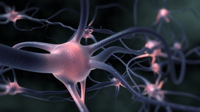
It’s not surprising but it is alarming: physical and emotional neglect has a harmful effect on children’s developing brains, new research shows.

Children’s Hospital Boston’s blog Vector pulls together several studies that detail the various levels of damage that can be done to children’s brains when they are subject to trauma, neglect and social isolation:
Sheridan, Nelson and colleagues obtained brain MRIs from three groups of 8- to 11-year old children: 29 had been reared in an institution, 25 had left the institution for a high-quality foster home (where they spent 6 to 9 years), and 20 typically developing children who were never institutionalized served as controls.

The findings were mixed:
–Children who had spent their entire lives in an institution had significantly lower volumes of white matter—necessary for making connections in the brain—in the cortex of the brain than did the controls. But if they were transferred into foster care, their white matter volume became indistinguishable from that in controls.

–Children spending any amount of time in an institution—even if later placed in foster care—had significantly smaller gray matter volumes in the cortex.
“White matter, which forms the ‘information superhighway’ of the brain, shows some evidence of ‘catch up,’” says Sheridan.
An earlier study in mice made the connection between neglect and social isolation and cognitive impairment on a cellular level. Again, from the Vector blog:
Earlier this month, the journal Science reported findings from the Boston Children’s Hospital lab of Gabriel Corfas, PhD, showing how neglect and social isolation lead to cognitive impairment at the cellular level.
The team, led by Corfas and postdoctoral fellow Manabu Makinodan, MD, PhD, simulated neglect in mice by placing them in isolation early in life.
The results were striking. Cells that make up the brain’s white matter, known as oligodendrocytes, failed to mature, and failed to produce proper amounts of myelin, the fatty “insulation” on nerve fibers that boosts the speed and efficiency of communication between different areas of the brain. This was especially true in the prefrontal cortex. The mice, for their part, showed reduced sociability and deficits in working memory.
Confirming the earlier research, Corfas’s team showed that the effects of social isolation are timing-dependent. If mice were isolated during a specific period in their development, they failed to recover sociability and memory function even when they were put back in a social environment. Conversely, if they were isolated after this so-called critical period, they remained normal.

Many female brains contain male DNA

brain neuron

In the first study of its kind, researchers have discovered that male DNA is commonly found in the brains of women – a finding that could hold important implications for diseases like Alzheimer’s disease and cancer.
Male DNA is likely transferred to female brains during pregnancy, according to researchers from the Fred Hutchinson Cancer Research Center in Seattle.  During this time, mothers and fetuses exchange and harbor genetic material and cells in a phenomenon called microchimerism.

This means, if a mother is pregnant with a boy, she will end up with male DNA in her system – potentially for the rest of her life. If the fetus is female, the mother will end up with genetic material from her daughter, though it is difficult for researchers to distinguish between two sets of female DNA in microchimerism studies.
What this means for treating diseases
Prior studies have observed fetal DNA in many other of the mother’s tissues and organs, but this is the first to confirm fetal cells can cross the blood-brain barrier and reside in the mother’s brain beyond pregnancy.
“We were interested in looking at the human brain because it’s never been looked at before, and it was really unknown if the cells of fetal origin could reach the brain,” study senior author Dr. Lee Nelson, a member of the Fred Hutchinson Cancer Research Center and professor of medicine at the University of Washington, told FoxNews.com.  Nelson and her colleagues performed autopsies on 59 brains of deceased females and detected male microchimerism in 63 percent of them.

Male microchimerism was distributed across multiple regions of the female brain, including those affected by dementia, and could persist for decades – potentially even an entire human lifespan.  According to the study, the oldest female with microchimerism detected in her brain was 94.

“The question naturally arises what role might the cells have in benefiting health and what role they play in diseases,” Nelson added.

The researchers hope further studies on microchimerism might shed new light on various diseases that affect the brain, such as Alzheimer’s, Parkinson’s or even brain tumors.

“These cells have access to the brain could help us understand different treatment options for diseases that aren’t well treated,” Nelson said.  “It’s a very exciting new area that opens up different possibilities, such as, what if these cells have anti-tumor potential?  For example, glioblastomas are deadly tumors, (which) have poor treatment options.  We’re very much in need of new potential options.”

The researchers also examined rates of Alzheimer’s disease in women with and without male microchimerism in their brains.  In previous studies, it has been suggested that Alzheimer’s may be more prevalent in women who have had children.  However, data from this study found the women who had been diagnosed with Alzheimer’s had lower rates, or concentrations, of microchimerism in their brains than those without dementia.

Studies of other tissues and organs have suggested both protective benefits and risks associated with male microchimerism in mothers.  In a study of breast cancer, male microchimerism appeared to play a protective role: Women with male DNA had a lower rate of the disease than those without.

But –  a colon cancer study showed the opposite: Mothers with microchimerism had higher rates of the disease.

Researchers have a theory behind the protective benefits of microchimerism: They believe the ‘outsider’ DNA may boost the capacity of the mother’s immune system to seek out and destroy antigens.

“The theory is because the cells persisting after pregnancy are half-genetically different, they have another small window to recognize antigens, or cells that are either malignant or pre-malignant,” Nelson said.  “They can look at the cells with a slightly different perspective.”

The researchers said the next steps are to further define the role of microchimerism in health and disease, and see if the fetal cells are able to differentiate into other cells and perform other functions in the mother’s body.

Military’s new binoculars read brain waves to find danger

brainwave binoculars.jpg 
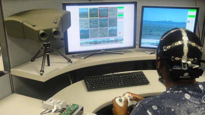
A brainwave-reading cap and a prototype binocular system combine to monitor threats far more effectively than a human acting alone.

By reading a soldier’s brain waves, new binoculars can detect a threat his brain has registered before he’s even conscious of it.

The Defense Advanced Research Projects Agency (DARPA) set out five years ago to provide the military with a far better device to visually detect threats. After all, the human eye’s limited field of view, breadth of area and the effects of fatigue can make relying on vision alone difficult.

They came up with the Cognitive Technology Threat Warning System program (CT2WS), binoculars paired that harness thoughts in the soldier’s brain.

It sounds like the stuff of science fiction, but CT2WS is based on real science: Humans are naturally skilled at detecting the out-of-the-ordinary -- and complex algorithms can interpret even subconscious human thoughts for computers to understand.

When the brain detects potentially threatening things like unexpected movements, it triggers a P-300 brain wave, a signal believed to connect with stimulus evaluation and categorization. The CT2WS system picks up those signals, translates them, and alerts the wearer of a threat through three key components.

The first is an electroencephalogram (EEG) cap that monitors the user’s brain signals. When the brain detects a threat, as seen through a tripod-mounted, 120-megapixel video camera with a 120-degree field of view, the cap records it. It's hardly the bird-watching binoculars you may be familiar with, but the system gets the job done.

The final part is complex visual processing algorithms that can run on a laptop, identifying potential targets and cuing images for the user to evaluate.

Testing revealed that while users were shown a rapid sequence of about ten images per second, their brains were still able to send signals flagging the relevant images, signals that could be read by the computer.

What about the classic rustle in the brush, however -- is it a bird or the enemy? The agency claims these cognitive algorithms have become so good that leaves rustling or wildlife movement that could be written off as typical nature will still be tagged as revealing potential threats.

Yet false alarm rates are greatly reduced through the EEG-based human filtering system.

Without a human and her brainwaves in the loop, the entire CT2WS system was tested and produced 810 false alarms per hour out of 2,304 events. But when a war fighter wears the EEG cap and contributes her brainpower to the system, false alarms were reduced to only 5 out of 2,304 events.

That’s a 91 percent improvement, thanks to those brain waves.

And target detection can reach 100 percent when commercial radar like Cerberus Scout is also deployed.
DARPA has been working with HRL Laboratories; Advanced Brain Monitoring; Quantum Applied Science & Research, Inc. and the University of California San Diego to create the system.

HRL Laboratories helped develop the sensor, cognitive and EEG decoding technologies. These sorts of advances are critical to harvesting an operator’s thought for the system.

Last year, the CT2WS was field-tested in a range of real environments including open desert at Arizona’s Yuma Proving Ground and tropical terrain in Hawaii, as well as open ground at California's Camp Roberts.
The results revealed the system can outperform trained humans, who detected about 60 percent of threats with binoculars. CT2WS wiped the floor with them, catching about 91 percent of risks.

Goals for this year included improving algorithms to increase frame rate and extending algorithms to handle imagery from Army and Marine Corps systems that generate visible, IR, and radar imagery from mast-mounted systems like Cerebus Scout.

Another critical aim for this year was to implement dry wearable EEG sensors that would not require conductive gels. DARPA awarded a contract to Quasar last September.

DARPA allocated $8.5 million last year and $1.75 million this year, providing a final demonstration to Army officials recently at Fort Belvoir, Va.

The CT2WS technology is currently being transitioned to the Night Vision and Electronic Sensors Directorate (NVEDS) in Fort Belvoir, where the first night vision goggles were created.

Since debuting those groundbreakers, NVEDS claimed to "own the night." Will they soon own the brain waves, too?