(HealthNewsDigest.com) - When your mind wanders, you're not paying attention to what's going in front of you. A new study suggests that it's not just the mind, it's the body, too; when subjects' minds wandered, they blinked more, setting up a tiny physical barrier between themselves and the outside world.
Cognitive neuroscientist Daniel Smilek, of the University of Waterloo, studies how people pay attention – and don't. For this study, he was inspired by brain research that shows, when the mind wanders, the parts of the brain that process external goings-on are less active. "And we thought, ok, if that's the case, maybe we'd see that the body would start to do things to prevent the brain from receiving external information," Smilek says. "The simplest thing that might happen is you might close your eyes more." So, Smilek and his colleagues, Jonathan S.A. Carriere and J. Allan Cheyne, also of the University of Waterloo, set out to look at how often people blink when their mind wanders.
Fifteen volunteers read a passage from a book on a computer. While they read, a sensor tracked their eye movements, including blinks and what word they were looking at. At random intervals, the computer beeped and the subjects reported whether they'd been paying attention to what they were reading or whether their minds were wandering – which included thinking about earlier parts of the text.
The participants blinked more when their minds were wandering than when they were on task, the team reports in Psychological Science, a journal of the Association for Psychological Science. "What we suggest is that when you start to mind-wander, you start to gate the information even at the sensory endings – you basically close your eyelid so there's less information coming into the brain," says Smilek.
This is part of a shift in how scientists are thinking about the mind, he says. Psychologists are realizing that "you can't think about these mental processes, like attention, separately from the fact that the individual's brain is in a body, and the body's acting in the world." The mind doesn't ignore the world all by itself; the eyelids help.
Psychological Science is ranked among the top 10 general psychology journals for impact by the Institute for Scientific Information.
Thursday, April 29, 2010
Human brains grow, change and can heal themselves
By the time Scott Hayner was 7, he had had one skull fracture and three major concussions from falling off horses.
Nobody connected those accidents to the difficulties he had in school. He acted out, stopped talking for three months and cried daily for two years. As an adult, he seemed to be a thriving, successful stockbroker, until traumatic brain injury from a 1999 soccer accident led to seizures and sidelined his ability to talk to people and stay on task, it seemed, for good.
Hope through brain plasticity
Two realizations have turned his life around at 42. First, he realized that brain injuries were behind the troubles he has had all his life. And second, he read about brain plasticity — the concept that the brain can heal and learn at all ages.
"It was a relief," says Hayner, who credits his 2008 training at the University of Texas at Dallas' Center for BrainHealth for helping to restore abilities that he thought were long gone. "It helped me regain my self-esteem and self-confidence. It gave me hope."
Neuroplasticity, or the brain's ability to adapt and change through life, is gaining increased traction in medical circles.
Dr. Norman Doidge, author of the best-selling "The Brain That Changes Itself: Stories of Personal Triumph from the Frontiers of Brain Science" (Penguin, $16), refers to neuroplasticity as "the most important change in our understanding of the brain in four hundred years.
"For the longest time our best and brightest neuroscientists thought of the brain as like a machine, with parts, each performing a single mental function in a single location," he wrote in an e-mail from the University of Toronto (he also teaches at Columbia University). "We thought its circuits were genetically hardwired, and formed, and finalized in childhood."
This meant that doctors assumed they could do little to help those with mental limitations or brain damage, he says — because machines don't grow new parts. The new thinking changes that: "It means that many disorders that we thought can't be treated have to be revisited."
Dr. Jeremy Denning, a neurosurgeon on the Baylor Plano medical staff, has seen that in his own
"The brain has the amazing ability to reorganize itself by forming new connections between brain cells," Denning says. "I have one patient I operated on a year ago who almost died from a hemispheric brain stroke and actually recovered from coma to hemiplegia (paralysis) to actually walking out of the hospital in four to five weeks. There are numerous studies looking at the changes that occur at the molecular level at the site of neuron connections. It is a very complex phenomenon, and we are still in the infancy of completely understanding it."
Lifelong adaptability
Dr. Sandra Chapman believes in lifelong plasticity. As founder of the Center for BrainHealth, she has set several studies in motion to explore how that concept can help those with brain damage and everyone else, including those with aging brains, middle-schoolers who need a brain boost and autistic children who need help rewiring the brain to improve their social cognition.
People such as Hayner have been able to benefit from some of these studies, although BrainHealth is primarily a research institute.
"Our brain is one of the most modifiable parts of our whole body," Chapman says.
That means that just as physical exercise keeps the body healthy, the right kind of learning will make it more likely for our brains to keep up with our ever-expanding life span, she notes.
Even while using the latest high-tech scanning devices to monitor results in her studies, when it comes to brain health, Chapman puts her greatest emphasis on a brain fitness exam that she refers to as a "neck-up checkup." It's done one-on-one using puzzles, paper, pen, pencil and just a few computer questions.
A "brain physical" at the center costs $600. Based on the results, experts recommend a simple, individualized strategy usually focusing on three key areas:
Strategic attention: the skill to block out distractions and focus on what's important. Exercises might include taking stock of your environment, identifying what distracts you and eliminating or limiting them, and creating daily priority lists.
•Integrated reasoning: the ability to find the message or theme in what you are watching, reading or doing. Exercises might include making a point of reflecting on the meaning of a book after you've read it or a movie after you've seen it and writing down your interpretation.
•Innovation: the vision to identify patterns and come up with new ideas, fresh perspectives and multiple solutions to problems. Exercises might include thinking of multiple solutions to problems as they come up, talking to other people to get a different perspective and taking time to step away from a problem to give yourself an opportunity for creative thoughts.
Hayner says his sessions — he attended for two months and completed take-home exercises — proved invaluable.
"I have been on so many drugs and medications, and they got me nowhere," he says. "Adults with TBIs (traumatic brain injuries) tend to become overwhelmed, and when someone becomes overwhelmed, it spirals into fear and chaos, and we have a tendency to shut down.
"Today as long as I stick to what I was taught here about filtering information and innovative thinking and what's important and what's not important and apply that to my real life, things don't confuse and baffle me. … I can make a decision on the important things that have to be done each day."
Things that can strain your brain
•Sleep deprivation
•Multitasking
•Stress
Concussion
•Some medications and sleep aids
•General anesthesia
•Failure to seek help if you notice difficulties such as loss of memory, inability to focus and make decisions, and a struggle to understand.
Source: the Center for BrainHealth
Sign Language Study Helps Explain How Human Brain Learns Language Unlike Any Other Species
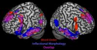
Instead, humans rely on several regions of the brain, each designed to accomplish different primitive tasks, in order to make sense of a sentence. Depending on the type of grammar used in forming a given sentence, the brain will activate a certain set of regions to process it, like a carpenter digging through a toolbox to pick a group of tools to accomplish the various basic components that comprise a complex task.
"We're using and adapting the machinery we already have in our brains," said study coauthor Aaron Newman. "Obviously we're doing something different [from other animals], because we're able to learn language unlike any other species. But it's not because some little black box evolved specially in our brain that does only language, and nothing else."
The team of brain and cognitive scientists – comprised of Newman (now at Dalhousie University after beginning the work as a postdoctoral fellow at the University of Rochester), Elissa Newport (University of Rochester), Ted Supalla (University of Rochester), Daphne Bavelier (University of Rochester), and Peter Hauser (Rochester Institute of Technology) - published their findings in the latest edition of the journal Proceedings of the National Academies of Science.
To determine whether different brain regions were used to decipher sentences with different types of grammar, the scientists turned to American Sign Language for a rare quality it has.
Some languages (English, for example) rely on the order of words in a sentence to convey the relationships between the sentence elements. When an English speaker hears the sentence "Sally greets Bob," it's clear from the word order that Sally is the subject doing the greeting and Bob is the object being greeted, not vice versa.
Other languages (Spanish, for example) rely on inflections, such as suffixes tacked on to the ends of words, to convey subject-object relationships, and the word order can be interchangeable.
American Sign Language has the helpful characteristic that subject-object relationships can be expressed in either of the two ways – using word order or inflection. Either a signer can sign the word "Sally" followed by the words "greets" and "Bob" (a construction in which word order dictates meaning), or the signer can use physical inflections such as moving hands through space or signing on one side of the body to convey the relationship between elements. For the study, the team formed 24 sentences and expressed each of those sentences using both methods.
Videos of the sentences being signed were then played for the subjects of the experiment, native signers who were lying on their backs in MRI (magnetic resonance imaging) machines with coils around their heads to monitor which areas of the brain were activated when processing the different types of sentences.
The study found that there are, in fact, distinct regions of the brain that are used to process the two types of sentences: those in which word order determined the relationships between the sentence elements, and those in which inflection was providing the information.
In fact, Newman said, in trying to understand different types of grammar, humans draw on regions of the brain that are designed to accomplish primitive tasks that relate to the type of sentence they are trying to interpret. For instance, a word order sentence draws on parts of the frontal cortex that give humans the ability to put information into sequences, while an inflectional sentence draws on parts of the temporal lobe that specialize in dividing information into its constituent parts, the study demonstrated.
"These results show that people really ought to think of language and the brain in a different way, in terms of how the brain capitalizes on some perhaps preexisting computational structures to interpret language," Newport said.
Aside from providing perspective on how language abilities might have evolved in humans, the scientists' findings could perhaps eventually find applications in medicine, according to Newport. For instance, it could prove valuable in assessing how best to teach language to a person with brain damage in certain areas but not others, such as a stroke victim.
How Exercise Might Help Keep Alzheimer's At Bay
People have been trying for years to keep their brains sharp by exercising, staying mentally active and watching their diets. But a panel convened by the National Institutes of Health warned the public Wednesday that it's not clear whether any of these measures can prevent Alzheimer's disease or other forms of mental decline in people.
Many scientists are still optimistic about prevention, partly because they are also considering research done on animals.
At about the time the panel was releasing its report, a 78-year-old senator was doing something he hopes is good for his brain.
Sen. Richard Lugar (R-IN) was competing in an annual charity race a few miles from Capitol Hill. He's been a runner since grade school and says he thinks exercise helps him remember a lot of stuff, including "the names and places of thousands of people and events that I bring up frequently in the course of debate.
"It's very helpful to have that kind of historical knowledge of my constituency, as well as of the world," Lugar said.
The panel convened by the NIH wasn't so sure that's why people like Lugar remain sharp into their 70s and 80s. And the panel wanted to make sure the public isn't being misled about the benefits of this or any other strategy for preventing Alzheimer's.
So panel members looked only at studies in humans, and they found that some studies of exercise in people have found a benefit while others haven't.
Arthur Kramer, a neuroscientist from the University of Illinois, wasn't on the panel, though he was invited to speak to the group.
He said the panel is right to be cautious, but that it also makes sense for researchers to talk about the potential of exercise.
"The benefits tend to be on the order of a 20 to 30 percent reduction in being diagnosed with Alzheimer's disease and other such diseases," Kramer said. "And again, this isn't universal but this is found in an increasing number of studies."
Kramer said researchers also tend to consider studies that show what exercise does for animals.
"There are improvements in the chemistry of the brain in terms of the molecules that protect the brain, increases in the number of connections between neurons, which allows us to encode new learning and memory, and even the birth of new neurons in one region of the brain that supports memory," he said.
Mental exercise is another strategy that seems like a good idea to many Alzheimer's researchers. After all, it appears to increase connections in the brain and perhaps make the brain more resilient — in animals and perhaps in people.
But Neil Buckholtz from the National Institute on Aging says the panel would need much more than that to recommend a specific activity to the public.
"Doing crossword puzzles, Sudoku, those kinds of things — they're interesting, but the evidence is not available at this point that they actually have an effect," Buckholtz said.
The panel seemed most skeptical about studies of drugs, diets and nutritional supplements.
Members found some evidence of benefit from omega-3 fatty acids like those in fish. But it found no convincing studies in people that antioxidants like vitamin E could make a difference.
Martha Clare Morris, a nutritional epidemiologist at Rush University Medical School in Chicago, is less skeptical. She says there is good evidence that some antioxidants work in animals.
"There's a very broad base of animal models showing that vitamin E protects the brain from neuron loss, from DNA damage, from oxidative damage," she said.
Panel members say they will consider changing their position on vitamin E and other popular prevention strategies when researchers show they work in people.
Brain cell study sheds light on epilepsy
PHILADELPHIA, April 29 (UPI) -- U.S. neuroscientists have identified a process that helps control the firing of neurons -- a finding they said sheds light on epileptic seizures.
The researchers from the Children's Hospital of Philadelphia and Tufts University School of Medicine in Boston said by isolating the molecular and electrical events that occur when the control is disrupted, the new research not only advances medicine's understand of epileptic seizures, but also potentially other diseases involving poorly regulated brain activity.
"By better understanding the detailed events that occur in epilepsy, we are gaining knowledge that could ultimately lead to better treatments for epilepsy, and possibly for other neurological diseases," said Children's Hospital neuroscientist Douglas Coulter, the study's corresponding author. "Temporal lobe epilepsy, in particular, often resists current treatments," he said.
Coulter's research group, collaborating with a Tufts University team led by co-senior author Professor Philip Haydon, explained that in epilepsy excessive signaling between neurons can lead to seizures. However, another class of brain cells called glia can regulate such signals. Among the glia are star-shaped cells called astrocytes -- the particular focus of this research.
"This study shows that changes in astrocytes are key to brain dysfunction and opens the potential for novel therapeutic strategies in epilepsy," Haydon said.
The study is detailed in the early online edition of the journal Nature Neuroscience.
Docs pull out 8cm splinter from boy's brain
NEW DELHI: The doctors were shocked when 11-year-old Rahul walked into the neurosurgery OPD at AIIMS a few days ago. Rahul had been walking about with an 8cm-long wooden splinter in his brain it had pierced his left eye for the past two years. On Wednesday, doctors at AIIMS extricated, in a five-hour-long surgery, the splinter from Rahul's brain. The doctors were very surprised that Rahul hadn't developed a neurological or other complication over the past two years.
"It's unbelievable. Despite having a foreign body in his brain, he didn't develop a medical complication. He was absolutely fine when he came to us,'' said Dr P Sarat Chandra, associate professor, AIIMS, who operated on Rahul.
According to Rahul's family, he was playing when he fell on a wooden stick, a splinter from which pierced his left eye and entered his brain. "The stick broke when he tried to get up. We took him to a local hospital where doctors said they couldn't remove it,'' said Ratna Devi, Rahul's mother, who took him to several hospitals before bringing him to AIIMS. Because of the accident, Rahul lost his vision in the left eye but that didn't stop him from going to school and going about his everyday activities.
Apart from the left eye orbit, the splinter pierced the temporal lobe (responsible for memory), cavernous sinus (a collection of thin-walled veins at the base of the brain), the cerebellum (responsible for motor function), and was exerting pressure on the brain stem. "The splinter missed the internal carotid artery, which is very close to the cavernous sinus, by less than a millimetre. Since the splinter had been lodged in the brain for two years, there was some inflammation around it and there were significant changes near the cerebellum. Had the splinter been pushed farther inside, it could have been fatal,'' said Chandra.
Doctors say Rahul's case is nothing short of a miracle. "All the major areas in the brain were affected by the splinter, but there were no complications. It is surprising that he didn't develop an infection over the past two years. And he is very lucky there was no major damage to the brain,'' said Dr AK Mahapatra, head of the department of neurosurgery, AIIMS.
During the surgery, doctors had to be very careful while pulling the splinter out. Even a minor error might have proved fatal. "We cut open the front of the left skull and the roof of the eye orbit. We retracted the brain so that we could see the splinter and then, slowly, pulled it out. The splinter had become soggy and it was difficult to pull it out in one piece. We then washed the affected areas of the brain with antibiotic solution,'' said Dr Chandra.
Doctors say that it will take Rahul a month or two to recover completely. "The risk of him catching an infection is high. He can still get meningitis so we have to be very careful,'' said Dr Mahapatra.
First McCullom Lake Brain Cancer Lawsuit Trials to Begin in June
 The first of more than two dozen toxic tort lawsuits filed on behalf of individuals diagnosed with brain cancer after living in the area of McCullom Lake, Illinois are set to go to trial in early June.
The McCullom Lake brain cancer lawsuits have been filed against various defendants, including Rohm and Haas, which operated a chemical plant in the area. While many defendants have settled out of court, the first trial against Rohm & Haas and other non-settling defendants is scheduled to begin in Philadelphia in a little over one month.
About 30 people out of a community of only 1,000 people have been diagnosed with various forms of brain cancer in recent years. The cancer rate in the community is extremely high, considering the national average is only 7 brain cancer cases per 100,000 people.
The number of brain cancer lawsuits for McCullom Lake residents has grown steadily since the first cases were brought in 2006.
Several of the cases were diagnosed through a $1.4 million cancer cluster class action lawsuit settlement reached with nearby Modine Manufacturing, Inc. in 2008. The original settlement fund has been used to provide numerous vouchers for pre-paid medical testing to past and present residents of the area to screen for brain cancer and brain tumors. At least two of the cases were detected through MRI scans performed for residents using the settlement medical vouchers.
Plaintiffs’ attorneys have said, despite the approaching trial, that there still could be more lawsuits filed as residents who have suffered injuries step forward.
The lawsuits contain allegations that Morton International and Rohm and Haas dumped chemicals illegally into local groundwater for decades, causing the unusually high cancer rate. Morton International owned the McCullom Lake chemical plant until 1999, when it was bought by Rohm and Haas. Rohm and Haas was purchased by Dow Chemical in April of last year.
Despite having admitted that chemicals were dumped in an eight-acre unlined pit for 20 years, Rohm and Haas has pledged to fight the lawsuits, saying their tests showed that contamination from the dumping flowed away from the village. In addition, the county tested 14 local wells and found no water contamination. However, plaintiffs contest the company’s claims about the flow of contaminants, and point out that the county tested the wells years after the company had ceased dumping, and never tested those wells while the groundwater was being contaminated.
Residents of another small town in Missouri have experienced a similar cluster of cancer, resulting in a number of brain tumor lawsuits for residents of Cameron, Missouri that allege the problems were caused by a tannery distributing toxic waste sludge to farmers for use as fertilizer on their fields. At least 70 Cameron, Missouri brain tumor cases have been diagnosed in that community of about 10,000 people.
Sex hormones control masculinization
Sex hormones controls ‘masculinization’ of the brain
A new study has uncovered some information about how sex hormones control masculinization of the brain during development and drive gender related behaviors in adult males.
Published by Cell Press in the April 29 issue of the journal Neuron , the study demonstrates that direct action of testosterone, the prototypical male hormone, is unnecessary for masculinizing the brain and behavior.
Testosterone and estrogen are thought to play an essential role in organizing and activating gender-specific patterns of behavior in sexually reproducing animals.
Testosterone is produced by the testes and directly activates the androgen receptor (AR) in target tissues such as muscle. Estrogen is produced by the ovaries and is nearly undetectable in the circulation of males of most species. However, circulating testosterone in males can be converted into estrogen in the brain, and this testosterone-derived estrogen has been shown to control many male behaviors.
"It was known that testosterone and estrogen are essential for typical male behaviors in many vertebrate species," explains the study’s senior author, Dr. Nirao M. Shah from the Department of Anatomy at the University of California, San Francisco. "However, how these two hormones interact to control masculinization of the brain and behavior remained to be established."
Dr. Shah and colleagues found that during the neonatal testosterone surge there is very little AR expressed in the developing brain, making it unlikely that testosterone signaling via AR plays a major role in masculinizing neural pathways. Importantly, they went on to show that the male pattern of AR expression in the brain was dependent on testosterone-derived estrogen signaling.
The researchers then used a genetic approach to knock out the AR in the mouse nervous system and observed that these mutants still exhibited male type mating, fighting, and territorial marking behaviours.
However, these mutant males had striking reductions in specific components of these masculine behaviors.
These results show that testosterone signaling via AR does not control masculine differentiation of the brain and behavior but regulates the frequency and extent of male typical behaviors.
"Our findings in conjunction with previous work suggest a model for the control of male pattern behaviors in which estrogen masculinizes the neural circuits for mating, fighting, and territory marking, and testosterone and estrogen signaling generate the male typical levels of these behaviors," concludes Dr. Shah. "It will be interesting in future studies to identify the molecular and circuit level mechanisms that are controlled by these hormones."
Supplements in fish oil do not boost brain power
Rejecting the long held belief that supplements in fish oil are good for kids brain, a new study claimed that the pills do not boost mental ability of children.
 For the study, which according to the researchers is the largest of its kind, 450 children aged eight to ten at 18 schools in South Wales were given either omega-3 supplements ‘clever capsules’ or placebos for a period of four months.
The results of a series of tests showed that the fish oil pills did not improve the youngsters’Supplements in fish oil do not boost brain power it did appear that those taking them were more attentive.
Further analysis showed that reading, spelling and handwriting were not improved in those who took omega-3, the Daily Mail reported.
Pointing that supplements might help some youngsters who have trouble concentrating in class, lead researcher Amanda Kirby said: “The primary message always has got to be to start with a good diet.”
Noting that kids are eating more rubbish, Kirby of the University of Wales, said: “If children have a relatively varied diet and don’t seem to have problems, it is probably not going to help them.”
For youngsters with attention deficit hyperactivity disorder or learning difficulties, fish oils are “worth a try,” she said.
She added: “Fatty acids make up 20 per cent of the brain and are going to have an effect in a number of different ways. Some of the studies on cardiovascular disease and Alzheimer’s disease are pretty convincing, but we need more research.”
Earlier studies have credited fats found in abundance in fish such as herring, mackerel, salmon and fresh tuna with health benefits from staving off heart disease, cancer and depression, to warding off Alzheimer’s disease.
Parkinson's eased by brain probe
 The invasive implant surgery is not suitable for all patients
 The research behind this news was a trial involving 366 people with advanced Parkinson’s disease that was not being adequately controlled with medication. It found that after a year, those who had a DBS implant had greater improvements in quality of life than those receiving medical treatment alone. This was particularly due to improvements in mobility, bodily discomfort and the ability to carry out the activities of daily living. However, DBS surgery was not without risks, and about 19% of patients had serious adverse effects, mainly infections.
This trial suggests that combining DBS with medication has some benefits beyond drug therapy alone. Importantly, though, DBS treatment is invasive and will not be appropriate for everyone with Parkinson’s. This means that the potential benefits of DBS would need to be balanced against its risks for each patient.
Where did the story come from?
This research was carried out by Professor Adrian Williams and colleagues from the Queen Elizabeth Hospital in Birmingham and other hospitals and research centres in the UK. The study was funded by the UK Medical Research Council, Parkinson’s UK and the Department of Health. It was published in the peer-reviewed medical journal, The Lancet.The BBC News website, Daily Mail and The Independent covered this story in an accurate and balanced way. The Daily Mail and BBC News reported that this was a decade-long trial, although the trial recruited participants between 2000 and 2006, so a number of the patients will not yet have been followed for a full ten years. The current results are also only based on follow-up in the year after surgery, with longer-term results awaited. The Independent reported that 5% of people receiving DBS had severe complications, such as infections. However, 19% were reported to have serious surgery-related adverse events in the research paper.
What kind of research was this?
This was a randomised controlled trial (RCT) called PD-SURG, which looked at the effect of deep brain stimulation (DBS) on quality of life in people with advanced Parkinson’s disease. Treatment with DBS involves implanting wire electrodes into the brain. These electrodes are attached to a “pacemaker” device, which regularly sends electrical impulses through the electrodes and into the brain. In most cases, the pacemakers in this trial were implanted into an area of the brain known as the subthalamic nucleus, although other DBS procedures may use alternative sites.An RCT is the most appropriate way to compare the effects of different treatments. This RCT compared the best medical treatment alone with the same type of medical treatment combined with a DBS implant. This study design would be the best way to tell whether DBS provided any additional benefits over and above standard treatment.
What did the research involve?
The researchers recruited 366 people with Parkinson’s disease that was not adequately controlled with medication alone. They were randomised to continue to receive best medical treatment alone (drugs such as dopamine agonists, MAO type B inhibitors, COMT inhibitors and apomorphine) or to receive DBS surgery in addition to the best medical treatment. The researchers followed the participants up for one year and measured their quality of life to see whether DBS had any effect on this outcome.The participants in this trial were enrolled at 13 neurosurgery centres in the UK between 2000 and 2006. They had to have Parkinson’s disease diagnosed according to standard criteria, and to be fit enough to undergo surgery. Before being randomised, the participants filled out a standard Parkinson’s disease questionnaire (PDQ-39), which assessed their quality of life. One year after being randomised and receiving their assigned treatment, the participants filled in this questionnaire again.
The researchers then compared changes in quality of life in the group that received DBS and the group that did not. A change of 10 points on the questionnaire score (based on a 39-point scale) was considered to be large enough to be meaningful to patients. A secondary outcome assessed by researchers was clinical assessment of the participants’ functioning using UPDRS scores, a standard scale for measuring Parkinson’s symptoms.
As one group had surgery and the other did not, it was not possible to blind participants to which treatment they received. The researchers also knew what treatments the participants had received as the study did not have sufficient resources to use independent blinded assessors for clinical assessments. People in the standard treatment group (the non-surgery group) could have surgery after one year if their treatment was still not adequately effective.
What were the basic results?
One year after surgery, people who received DBS in addition to best medical treatment showed greater improvement in their quality of life than those who received best medical treatment alone. The DBS group improved by 5 points on the PDQ-39 scale and the medical group by only 0.3 points.The quality of life questionnaire assessed different areas of life and showed that people who received DBS had greater improvements in mobility, activities of daily living and bodily discomfort. The difference between the groups was 8.9 points for mobility, 12.4 points for activities of daily living, and 7.5 points for bodily discomfort. Participants who received DBS also showed greater improvements in clinically assessed overall functioning at one year than participants receiving medication alone. Participants who received DBS had reduced their drug dose by about 34% compared with the medical treatment group.
Just under one in five people who received DBS had serious adverse effects associated with their surgery (19%), and one patient died from bleeding during surgery. Similar proportions of patients had side effects of their medical treatment in both groups (11% with DBS plus medical treatment, and 7% with medical treatment alone).
How did the researchers interpret the results?
The researchers concluded that one year after the study began, treatment that combined surgery and best medical therapy “improved patient self-reported quality of life more than best medical therapy alone in patients with advanced Parkinson’s disease”.They also say that the improvements seen were clinically meaningful, but that the risks associated with DBS surgery may warrant only offering the surgery to those people most likely to benefit from it.
Conclusion
This study used a robust design to assess the effects of deep brain stimulation (DBS) on quality of life in people with Parkinson’s disease that had not responded adequately to medical treatment. Points to note include:- Blinding participants and researchers to the treatment received was not possible, so participants’ ratings of their quality of life may have been affected if they had pre-existing expectations of DBS or if they were disappointed not to have received DBS.
- The trial has so far collected and reported one year’s worth of data. The researchers are continuing to collect information on the patients’ outcomes so that the longer-term effects of DBS can be studied.
- The researchers suggest that the group of patients treated were representative of those who would be offered surgery at neuroscience centres in the UK.
- A questionnaire was given to participants in the DBS group about surgery-related adverse effects six months after surgery, but a similar questionnaire was not given to the medical treatment only group. Therefore, adverse effects in the latter group could have been missed. The researchers also note that they did not record adverse effects that were not serious enough to cause a patient to be admitted to hospital or to extend their stay in hospital.
- People who received DBS continued to receive medical therapy, although the drug dose could be reduced in many cases. Therefore, news reports that “brain surgery is more effective than medication” or “implants have given us our life back” should not be misinterpreted to mean that DBS is a complete cure or that a person will no longer need any form of drug treatment. People should also be aware that all surgical procedures are associated with some degree of risk and this treatment would not be suitable for everyone. Advances and developments in the DBS technique are likely to continue.
Health Matters: Working Out Boosts Brain Power
A new study says working out is not only good for your body but also your brain.
An aerobic workout is more beneficial than you might think. It is now credited with helping to boost brain power.
Researchers at the University of Pittsburgh tested two groups of female monkeys.
One group ran on a treadmill for one hour each day, five days per week, for five months.
And the other - remain sedentary.
Their findings -- monkeys who exercised regularly at an intensity that would improve a human's fitness actually boosted their learning power and had greater blood volume to their brains.
Researchers say the same results can be found in humans - because of our close physiology to monkeys.
The findings are in the journal Neuroscience.
Subscribe to:
Comments (Atom)