Friday, April 2, 2010

Leave Your Biceps & Start Flexing All Your Brain Muscles To Get Through This Mindflex Game

Science is still confused when it comes to telekinesis because it’s something which many believe in but hardly anyone has seen it being done, and people are so curious to know about it that a person called James Randi who’s a well known rich disbeliever of all these paranormal things, announced a prize money of $1,000,000 to show him so telekinesis in reality.
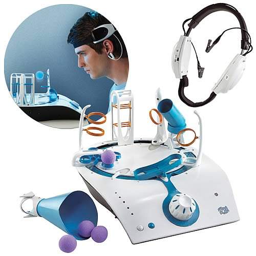
I think with this one, you can attempt to take away the prize money of 1 million. Here is “Mindflex game” which flexes your brain muscles to move the objects. Actually it just gives an example of how powerful the brain is and can be if it’s enhanced. This mental acuity game will surely open the eyes of all telekinesis skeptics.
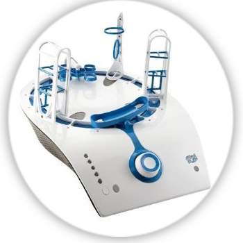
It’s coming with a lightweight headset which has been connected to small sensors for forehead and earlobes and works in quite an interesting way. I feel you might just laugh at the following sentence but being serious and writing no jokes, it actually measures your brainwave activity. Your concentration level determines how much power your brain can exert and thus leading to the creation of a small foam ball. The more you concentrate, the more this ball will rise gently on a stream of air. Once you relax your thoughts and voila, the ball will descend.
Using your mental and physical coordination, you will have to guide it through various obstacle courses.  Being for players of ages 8 and up, it’s a killer game and tests how much your brains can handle. If you want to buy it then order it for $99.99. It will be available in March, and a demo video can be found below.

Pfizer, Onyx drug offers hope against brain cancer

Researchers from Georgetown Lombardi Comprehensive Cancer Center and the University of California said the experimental drug, called PD-0332991, may become a new treatment option for glioblastoma, the commonest and deadliest form of brain cancer.

Clinical trials to test the drug in patients with recurrent brain cancer are under development, they said in a study published in the journal Cancer Research.

“We don’t know how well this agent will perform in patients with glioblastoma but in the mice we studied we saw very impressive, durable effect,” said David James, a professor of neurological surgery at UCSF, who worked on the study.

“What is especially encouraging about this agent is that we found it can easily pass through the blood-brain barrier and access glioblastoma, and that there is already a simple test available for screening glioblastoma patients in advance to see whether or not they should be responsive to this therapy.”

Data from a recently-published study by The Cancer Genome Atlas Research Network, suggests about 90 percent of glioblastoma patients would be suitable candidates for the drug, he said.

PD-0332991, which Pfizer is developing under license from Onyx, is being tested in human trials for other cancers such as multiple myeloma and mantle cell lymphoma. It is a pill designed to shut down activity of molecules called cyclin-dependent kinases 4 and 6 (cdk4 and cdk6) that drive cell division.

“In normal cells these kinases are kept under exquisite control by a gene known as p16,” said Todd Waldman of Lombardi, who worked on the study with James.

“But in glioblastoma and other cancers, p16 is frequently deleted, and these two kinases are uncontrollably activated, which drives the cell to divide and form cancer.”

But the drug does not work if the cancer is missing a protein known as retinoblastoma (Rb). A test for Rb is already being used to screen patients for use of PD-0332991 in the ongoingclinical trials.

In their study James’s team implanted three different kinds of human glioblastoma directly into the brains of mice and then treated them with PD-0332991. They found the drug was able to get to the tumors and halt the cancer’s growth as long as the mice stayed on the drug.

Because PD-0332991 itself does not kill cancer cells — just halts their growth — the researchers then combined the drug with radiation and found the combination worked better than PD-0332991 alone. They also successfully tested the drug in mice in which glioblastoma had come back after treatment with temozolomide, a chemotherapy used in many cancer patients.

Anxiety modifies depression's ill effects

Anxiety may modify depression for better or for worse, says a new study.

The research, thereby, establishes a link between anxiety and depression, claiming that they co-occur.

The study, in the journal Cognitive, Affective & Behavioral Neuroscience , looked at depression and two types of anxiety: anxious arousal, the fearful vigilance that sometimes turns into panic; and anxious apprehension, better known as worry.

In their research, boffins used functional Magnetic Resonance Imaging (fMRI) at the Beckman Institute’s Biomedical Imaging Center to look at brain activity in subjects who were depressed and not anxious, anxious but not depressed, or who exhibited varying degrees of depression and one or both types of anxiety.

"Although we think of depression and anxiety as separate things, they often co-occur," said University of Illinois psychology professor Gregory A. Miller, who led the research with Illinois psychology professor Wendy Heller. "In a national study of the prevalence of psychiatric disorders, three-quarters of those diagnosed with major depression had at least one other diagnosis. In many cases, those with depression also had anxiety, and vice versa."

In the study, brain scans were done while participants performed a task that involved naming the colors of words that had negative, positive, or neutral meanings. This allowed the researchers to observe which brain regions were activated in response to emotional words.

The researchers found that the fMRI signature of the brain of a worried and depressed person doing the emotional word task was very different from that of a vigilant or panicky depressed person.

"The combination of depression and anxiety, and which type of anxiety, give you different brain results," Miller said.

Perhaps most surprisingly, anxious arousal (vigilance, fear, panic) enhanced activity in that part of the right frontal lobe that is also active in depression, but only when a person’s level of anxious apprehension, or worry, was low. Neural activity in a region of the left frontal lobe, an area known to be involved in speech production, was higher in the depressed and worried-but-not-fearful subjects.

Despite their depression, the worriers also did better on the emotional word task than those depressives who were fearful or vigilant. The worriers were better able to ignore the meaning of negative words and focus on the task, which was to identify the color – not the emotional content – of the words.

Japanese Encephalitis: Inflammation of the Brain

Japanese encephalitis (JE) is an inflammation of the brain, due to infection by a virus known as a flavivirus. Occuring most commonly in Southeast Asia and the Far East and the Pacific islands, this infection in humans is the result of a mosquito bite.

Pigs and wild birds are the initial carriers, who pass it on to mosquitoes. People do not catch this virus from other people.

In most cases, symptoms are relatively mild, often no more than fever, loss of appetite, and headache. Dizziness, nausea, vomiting and diarrhea may also occur. The effects are similar to a case of the flu.
Click here to comment on this article.

In more serious infections, the onset is sudden, with headache, stiff neck, high fever, sweating and tremors. Breathing may be rapid and shallow. Meningitis (infection of the meninges, which are membranes surrounding the brain and spinal cord) will often develop.

The individual may be gripped by convulsions or paralysis. They may be disoriented, and ultimately lapse into a coma.

Inflammation of the brain develops in about one out of every 500 cases. Progression of the disease often culminates in brain damage or even death.

Many of those fortunate enough to recuperate, discover that recovery is rarely total. The individual usually will carry some degree of disability either mentally or physically.

They may continue to experience lack of muscle coordination (ataxia), seizures, paralysis, as well as neurological and cognitive (thinking) impairment.

However, in the many mild cases of this disease, the individual will usually become immune to the virus.

The incubation period for JE is between five and 15 days after being bitten by an infected mosquito. Within another five to 15 days, symptoms begin to appear. They will last between one and six days.

JE, also known as Japanese B encephalitis, is the most common viral encephalitis that can be transmitted to human beings. It is also the main cause of viral encephalitis in Asia. More than 30,000 cases are reported every year.

The people most likely to be affected by this virus live in rural areas. U.S. civilians, and those in the military in Asia, make up only one case per year.
All available treatments can do little more than attempt to keep symptoms from worsening. Possible treatments are antiviral drugs, cortisone, anti-seizure medication, and acetaminophen for fever and headache.

In extreme cases, hospitalization may be in order, where the goal will be to reduce pain and fever.

Behavioral incentives mimic effects of medication on brain systems in ADHD

Medication and behavioural interventions help children with Attention Deficit Hyperactivity Disorder (ADHD) better maintain attention and self control by normalising activity in the same brain systems, according to research funded by the Wellcome Trust.

In a study published today in the journal Biological Psychiatry, researchers from the University of Nottingham, show that medication has the most significant effect on brain function in children with ADHD, but this effect can be boosted by complementary use of rewards and incentives which appear to mimic the effects of medication on brain systems.

ADHD is the most common mental health disorder in childhood, affecting around one in twenty children in the UK. Children with ADHD are excessively restless, impulsive and distractible and experience difficulties at home and in school. Although no cure exists for the condition, symptoms can be reduced by a combination of medication and behaviour therapy.

Methylphenidate, a drug commonly used to treat ADHD, is believed to increase levels of dopamine in the brain. Dopamine is a chemical messenger associated with attention, learning and the brain's reward and pleasure systems. This change amplifies certain brain signals and can be measured using an electroencephalogram (EEG). Until now it has been unclear how rewards and incentives affect the brain, either with or without the additional use of medication.

To answer these questions, researchers at the university's 'Motivation, Inhibition and Development in ADHD Study' (MIDAS) used EEG to measure brain activity whilst children played a simple game. They compared two particular markers of brain activity that relate to attention and impulsivity and looked at how these were affected by medication and motivational incentives.

The team worked with two groups of children aged nine to fifteen years, one group of twenty-eight children with ADHD and a control group of twenty-eight children. The children played a computer game in which green aliens were randomly interspersed with less frequent black aliens, each appearing for a short interval. Their task was to 'catch' as many green aliens as possible, while avoiding catching black aliens. For each slow or missed response, they would lose one point; they would gain one point for each timely response.

In a test designed to study the effect of incentives, the reward for avoiding catching the black alien was increased to five points; a follow-up test replaced this reward with a five point penalty for catching the wrong alien.

The researchers found that when given their usual dose of methylphenidate, children with ADHD performed significantly better at the tasks than when given no medication, with better attention and reduced impulsivity. Their brain activity appeared to normalise, becoming similar to that of the control group.

Similarly, motivational incentives also helped normalise brain activity on the two EEG markers and improved attention and reduced impulsivity, though its effect was much smaller than that of medication.

"When the children were given rewards or penalties, their attention and self control was much improved," says Dr Maddie Groom, first author of the study. "We suspect that both medication and motivational incentives work by making a task more appealing, capturing the child's attention and engaging his or her brain response control systems."

Professor Chris Hollis, who led the study, believes the findings may help reconcile the often-polarised debate between those who advocate either medication on the one hand, or psychological/behavioural therapy on the other.

"Although medication and behaviour therapy appear to be two very different approaches of treating ADHD, our study suggests that both types of intervention may have much in common in terms of their affect on the brain," he says. "Both help normalise similar components of brain function and improve performance. What's more, their effect is additive, meaning they can be more effective when used together."

The researchers believe that the results lend support from neuroscience to current treatment guidelines for ADHD as set out by the National Institute for Health and Clinical Excellence (NICE). These recommend that behavioural interventions, which have a smaller effect size, are appropriate for moderate ADHD while medication, with its larger effect size, is added for severe ADHD.

Although the findings suggest that a combination of incentives and medication might work most effectively, and potentially enable children to take lower doses of medication, Professor Hollis believes more work is needed before the results can be applied to everyday clinical practice or classroom situations.

"The incentives and rewards in our study were immediate and consistent, but we know that children with ADHD respond disproportionately less well to delayed rewards," he says. "This could mean that in the 'real world' of the classroom or home, the neural effects of behavioural approaches using reinforcement and rewards may be less effective."

Inside the Science of Laughter

This April Fools' Day, regardless of whether you're the one playing the prank or the victim of someone else's shenanigans, odds are you'll be laughing--and to Baltimore based neuroscientist Robert Provine, that's serious business.

Provine, author of the book "Laughter: A Scientific Investigation" and the subject of a March 31 article by Associated Press (AP) science writer Seth Borenstein, believes that only 10-percent to 15-percent of a person's laughter comes as the result of jokes or riddles. Rather, he says, "Laughter above all else is a social thing… The requirement for laughter is another person."

The University of Maryland Baltimore County professor has spent years scientifically dissecting humor and its response. He says that each laugh lasts approximately 1/15th of a second, and is repeated every 1/5th of a second. Additionally, it is not reliant on any single sense, as people can and often do laugh without seeing or hearing a specific trigger.

Also, Provine says, laughter knows know linguistic boundaries. "All language groups laugh `ha-ha-ha' basically the same way," he told Borenstein. "Whether you speak Mandarin, French or English, everyone will understand laughter… There's a pattern generator in our brain that produces this sound."

Like Provine, Bowling Green State University (BGSU) psychology professor Jaak Panksepp has studied the science of laughter--in particular, laughter amongst animals such as chimpanzees and rats. Panksepp has documented his research, in which he tickles rats and receives a laugh in response, on YouTube and in various scientific journals.

According to Borenstein, the BGSU professor and others like him hope to "figure out what's going on in the brain during laughter. And it holds promise for human ills… Northwestern University biomedical engineering professor Jeffrey Burgdorf has found that laughter in rats produces an insulin-like growth factor chemical that acts as an antidepressant and anxiety-reducer. He thinks the same thing probably happens in humans, too. This would give doctors a new chemical target in the brain in their effort to develop drugs that fight depression and anxiety in people."

Perhaps what they say is true--maybe laughter really is the best medicine.

Magnets Can Alter Moral Judgement By Changing Brain Activity

US scientists have discovered that appyling a magnetic field to a particular place on the scalp can alter people's moral judgement by interfering with activity in the right temporo-parietal junction (TPJ) of the brain. They said their finding helps us better understand how the brain constructs morality.

You can read about the study, led by researchers from the Massachusetts Institute of Technology (MIT), in Cambridge, Massachusetts, in the 29 March online issue of the Proceedings of the National Academy of Sciences, PNAS. The research was led by Dr Rebecca Saxe, assistant professor of brain and cognitive sciences at MIT.

Lead author Dr Liane Young, a postdoctoral associate in Saxe's department, told the media that because people are normally very confident and consistent in making moral judgements, it comes as surprise to learn that their ability to do so can altered like this.

"You think of morality as being a really high-level behavior. To be able to apply (a magnetic field) to a specific brain region and change people's moral judgments is really astonishing," said Young in a statement.

She said the study reveals "striking evidence" that the right TPJ, which sits on the surface of the brain, above and behind the right ear, plays a crucial role in making moral judgements.

When we make moral judgements about other people we often need to infer their intentions. For instance, when a hunter on a hunting trip shoots a fellow hunter, did he mistake his colleague for prey, or was he secretly jealous?

This ability has been termed "theory of mind", that is the ability to attribute mental states such as beliefs, intentions, and other qualities to oneself and others, and also to understand that other people's mental states can be different to one's own.

Ten years ago Saxe identified that the TPJ played a role in theory of mind and wrote about it in her PhD thesis in 2003. Since then she has been using functional magnetic resonance imaging (fMRI) to show that the right TPJ is active when people are asked to make moral judgements that require them to think about the intentions of others.

Other studies have also shown that the TPJ is highly active when we think about other people's intentions, their beliefs and their thoughts.

For this study, Saxe, Young and colleagues wanted to investigate what might happen if they could actually disrupt activity in the right TPJ.

In this case, instead of the usual fMRI, they did two sets of experiments where they used a non-invasive method called transcranial magnetic stimulation (TMS) to apply a magnetic field to a small area of the skull (on the scalp) to create weak electric currents that stop nearby brain cells from firing normally for a while.

They found that this was enough to impair subjects' ability to make moral judgments that involve an understanding of other people's intentions: as in for example, a failed murder attempt.

In the first set of "offline stimulation" experiments, they exposed volunteers to the TMS method for 25 minutes and then asked them to take a test where they read about several scenarios and then had to judge the actions of the characters portrayed on a scale of one to seven (from "absolutely forbidden" to "absolutely permissible").

For example, for one scenario they were asked to judge how permissible would it be for a man to allow his girlfriend to walk across a bridge he knew to be unsafe, even if she does eventually cross it safely. In such a scenario, judging the man solely on the outcome would hold him blameless, even though he apparently intended harm.

In the second set of "online stimulation" experiments, the volunteers underwent a 500-millisecond burst of TMS at the point when they were asked to make a moral judgement.

In both experiments, Saxe, Young and colleagues found that disrupting the right TPJ resulted in volunteers being more likely to judge failed attempts to harm as morally permissible.

They suggested this was because they were relying more on information about the outcome than inference on intention, since the process that normally helped them get information on intention was disrupted by the electrical current from the TMS.

"It doesn't completely reverse people's moral judgments, it just biases them," explained Saxe.

The researchers also found that when they applied TMS to a brain region near the right TPJ , the volunteers' judgments were nearly identical to those of volunteers who received no TMS at all.

They concluded that:

"Relative to TMS to a control site, TMS to the RTPJ caused participants to judge attempted harms as less morally forbidden and more morally permissible. Thus, interfering with activity in the RTPJ disrupts the capacity to use mental states in moral judgment, especially in the case of attempted harms."

When we judge other people, understanding their intentions is just one aspect of what we take into account. We also assess things like their previous record, what we understand about their desires, and what constraints they might be under. We are also guided by our own ideas about loyalty, fairness and integrity, said Saxe.

Moral judgement is not a single process, even though it might feel like it, explained Saxe, who described it as more a mixture of "competing and conflicting judgments, all of which get jumbled into what we call moral judgment".

Dr Walter Sinnott-Armstrong, professor of philosophy at Duke University, who was not involved in this research, said that by going beyond fMRI, the study marks a major step forward for the field of moral neuroscience:

"Recent fMRI studies of moral judgment find fascinating correlations, but Young et al usher in a new era by moving beyond correlation to causation," said Sinnott-Armstrong.

The National Center for Research Resources, the MIND Institute, the Athinoula A. Martinos Center for Biomedical Imaging, the Simons Foundation and the David and Lucille Packard Foundation funded the study.