Monday, November 19, 2012

Four-Year-Old Brain Cancer Patient

Cancer claimed the life of medical marijuana poster child Cash “Cashy” Hyde after access to cannabis-based medication was cut off by a sweeping medical marijuana crackdown in Montana last summer

MISSOULA, MT — Cash “Cashy” Hyde passed away in his father’s arms this week. The four-year-old Montana boy—whose malignant brain cancer had been in remission while he received consistent medical cannabis treatments—is the latest casualty in the federal government’s war on voter-approved medical marijuana, says John Malanca, a close friend of the Hyde family.

Malanca recounts the Hyde family’s ordeal:
Desperate to find anything to keep their young child Cash alive, after traditional treatments and medications failed to slow the growth of the malignant tumor in his brain, Mike and Kalli Hyde turned to high-CBD cannabis oil. Cash’s cancer immediately went into remission, and he started to live a more normal life.
Cash was comfortable, started eating again, and recovered his desire to play.

Then, last summer, law enforcement officials in Montana came down hard on the medical marijuana industry.
Rather than face the risk of being stormed by armed agents and subjected to steep fines or jail time, many legally-compliant medical cannabis dispensaries in Montana closed down.

The federal crackdown resulted in the closure of most of the medical marijuana dispensaries in Montana, and cut off the Hyde’s access to the therapeutic cannabis oil that was keeping Cash’s cancer at bay. Once Cash’s medication was cut off, his cancer came back out of remission.

“We put out a call to action, asking people to donate cannabis oil for Cash’s health—and there was a heart-warming outpouring of donors and support,” said Malanca. “Unfortunately, by the time we got Cash the medication he needed, his cancer could no longer be controlled.”

The Hyde Family has established the Cash Hyde Foundation to help prevent unnecessary loss of health and life through advocacy of progressive and responsible medical marijuana policies, law enforcement, and legislation: “Cashy’s Law.”

The foundation also supports increased medical marijuana research, cannabis health education, and pediatric cancer research.

“Cannabis oil extended Cash’s life; he beat cancer twice,” said Malanca. “He brought so much love to this world and so many people together. He educated people.”

Effect Of Trance-like States On The Brain Studied Using Brazilian Mediums


In this week’s online open source journal PLOS ONE, a new study details how brain activity affects individuals who engage in psychography. Psychography, or automatic writing, is a technique used by mediums in an effort to free-write messages from the deceased or from spirits.

Researchers from Thomas Jefferson University and the University of Sao Paulo in Brazil embarked on this study, analyzing the cerebral blood flow (CBF) of Brazilian mediums during this mystical practice. What the team was able to determine was that the brain activity of the mediums underwent a significant decrease in activity when they would enter this mediumistic dissociative state.

To collect the data, researchers selected 10 mediums and injected them with a radioactive tracer that would allow them to visualize their brain activity during both normal writing and also during the practice of psychography. Of the 10 mediums observed in the study, five were considered as experienced while the other five were less expert. To observe the brain activity, researchers employed the use of SPECT (single photon emission computed tomography), allowing them to conclusively view the brain and the areas within it that were active and inactive at different times during the experiment.

“Spiritual experiences affect cerebral activity, this is known. But, the cerebral response to mediumship, the practice of supposedly being in communication with, or under the control of the spirit of a deceased person, has received little scientific attention, and from now on new studies should be conducted,” says Andrew Newberg, MD, director of Research at the Jefferson-Myrna Brind Center of Integrative Medicine and a nationally-known expert on spirituality and the brain, who collaborated with Julio F. P. Peres, Clinical Psychologist, PhD in Neuroscience and Behavior, Institute of Psychology at the University of Sao Paulo in Brazil, and colleagues on the research.

With between 15 and 47 years of automatic writing experience, and having performed the act as many as 18 times a month, each of the mediums was also right-handed, found to be in satisfactory mental health and not currently using any type of psychiatric drugs. Each medium was able to report that during the study, the trance-like state associated with psychography was achieved and that during the normal writing control task each was in a regular state of consciousness.

The data collected from both the experienced and non-expert mediums showed two different outcomes. The researchers noted that while the experienced psychographers showed a lowered level of activity, the less-expert psychographers showed an increase in CBF in the same observed area. The area that the SPECT focused on was the left hippocampus (limbic system), right superior temporal gyrus, and the frontal lobe regions of the left anterior cingulated and right precentral gyrus during both the act of psychography as compared to their normal, non-trance writing. This region has been deemed to be important due to its association with reasoning, planning, generating language, movement and problem solving. The lowered activity for the experienced mediums suggests, according to researchers, an absence of focus, self-awareness and consciousness during psychography.

As mentioned, the less expert psychographers presented data that showed the opposite from their more experienced counterparts. Their increased levels of CBF in the same frontal areas may be related to their more purposeful attempt at performing psychography.

The team pointed out that as none of the mediums had current mental disorders, their data supports currently held evidence that dissociative experiences are not uncommon in the general population and are not necessarily indicative of a mental disorder, especially when experienced in a religious or spiritual context. 
They do believe that additional research should be conducted to specifically address criteria for distinguishing between both healthy and pathological dissociative expression as it relates to mediumship.

The team also performed a detailed analysis of the writing samples that were collected. What they determined was that the complexity scores for the trance-induced writing was much higher than the control writing was. The more experienced mediums showed the highest complexity scores, which one would think would actually require more CBF activity in the frontal and temporal lobes. The writings composed during psychography typically involved ethical principles, the importance of spirituality, and bringing together the fields of science and spirituality.

Researchers have developed a few different hypotheses for why they think their data showed what it did. The first states that as frontal lobe activity decreases, the areas of the brain that support mediumistic writing are further disinhibited so that the overall complexity can increase. This process is similar to what is seen during alcohol and drug use. According to Newberg, “While the exact reason is at this point elusive, our study suggests there are neurophysiological correlates of this state.”

“This first-ever neuroscientific evaluation of mediumistic trance states reveals some exciting data to improve our understanding of the mind and its relationship with the brain. These findings deserve further investigation both in terms of replication and explanatory hypotheses,” states Newberg.

Brain power

Taking the pill to ward off dementia is not advised, says Arlene Harris. THE contraceptive pill has been lambasted and heralded since its conception in the 1960s. Some studies have found it to be a possible cause of cancer, while others have claimed it helps to prevent it. But it hasn’t been linked to brain health — until now.

Scientists at the University of Wisconsin have suggested that women in their 50s who took the pill during their earlier years performed better in memory tests than those who had never taken it and, in fact, may be less likely to develop dementia in later life.

The American researchers believe that oestrogen — which is the main hormone found in the pill — prevents hardening of the arteries, which increases blood supply to the brain and in turn helps to stave off dementia.

Lead researcher Kelly Egan, whose study is published in the Journal of Women’s Health, said: "Our analysis indicated that hormonal contraceptive use may have a protective cognitive (memory) effect, even years after use is discontinued. This is especially true in subjects with a longer duration of use."

But the Irish Medicines Board says this research is not conclusive and people would be advised not to start taking the pill for the sole purpose of preventing dementia.

"The IMB is aware of this study carried out by the University of Wisconsin," says a spokeswoman. "The authors themselves acknowledge the many limitations of the study and advise caution against interpretation of their results.

"Much more research is necessary in larger more focussed clinical trials before any conclusions can be made on this hypothesis."

Dr Shirley McQuade of the Dublin Well Woman Clinic agrees: "The reasons for dementia are complex and multifactorial," she says. "There probably is a link to oestrogen because previous studies have shown women who go through an early surgical menopause due to removal of their ovaries are more likely to have some cognitive impairment such as increased levels of forgetfulness.

"There have also been a number of studies looking at the possible protective effect of HRT on the development of dementia. However, it’s a bit of a leap from there to saying the contraceptive pill might be protective against dementia in the vast majority of normal women."

Matthew Gibb, senior social worker at the Memory Clinic in St James’ Hospital, Dublin, says looking after our vascular health will help to prevent hardening of the arteries.

"We would always advise people that what is good for the heart is good for the brain," he says.

Clues to Cause of Kids' Brain Tumors

TEHRAN (FNA)- Insights from a genetic condition that causes brain cancer are helping scientists better understand the most common type of brain tumor in children.
In new research, scientists at Washington University School of Medicine in St. Louis have identified a cell growth pathway that is unusually active in pediatric brain tumors known as gliomas. They previously identified the same growth pathway as a critical contributor to brain tumor formation and growth in neurofibromatosis-1 (NF1), an inherited cancer predisposition syndrome.

"This suggests that the tools we've been developing to diagnose and treat NF1 may also be helpful for sporadic brain tumors," says senior author David H. Gutmann, MD, PhD, the Donald O. Schnuck Family Professor of Neurology.

The findings appear Dec. 1 in Genes and Development.

NF1 is among the most common tumor predisposition syndromes, but it accounts for only about 15 percent of pediatric low-grade gliomas known as pilocytic astrocytomas. The majority of these brain tumors occur sporadically in people without NF1.

Earlier research showed that most sporadic pilocytic astrocytomas possess an abnormal form of a signaling protein known as BRAF. In tumor cells, a piece of another protein is erroneously fused to the business end of BRAF.

Scientists suspected that the odd protein fusion spurred cells to grow and divide more often, leading to tumors. However, when they gave mice the same aberrant form of BRAF, they observed a variety of results. Sometimes gliomas formed, but in other cases, there was no discernible effect or a brief period of increased growth and cell division. In other studies, the cells grew old and died prematurely.

Gutmann, director of the Washington University Neurofibromatosis Center, previously showed that mouse NF1-associated gliomas arise from certain brain cells.

According to Gutmann, the impact of abnormal NF1 gene function on particular cell types helps explain why gliomas are most often found in the optic nerves and brainstem of children with NF1 -- these areas are where the susceptible cell types reside.

With that in mind, Gutmann and his colleagues tested the effects of the unusual fusion BRAF protein in neural stem cells from the cerebellum, where sporadic pilocytic astrocytomas often form, and in cells from the cortex, where the tumors almost never develop.

"Abnormal BRAF only results in increased growth when it is placed in neural stem cells from the cerebellum, but not the cortex," Gutmann says. "We also found that putting fusion BRAF into mature glial cells from the cerebellum had no effect."

When fusion BRAF causes increased cell proliferation, postdoctoral fellows Aparna Kaul, PhD and Yi-Hsien Chen, PhD, showed that it activates the same cellular growth pathway, called mammalian target of rapamycin (mTOR), that is normally also controlled by the NF1 protein. An extensive body of research into the mTOR pathway already exists, including potential treatments to suppress its function in other forms of cancer.

"We may be able to leverage these insights and our previous work in NF1 to improve the treatment of these common pediatric brain tumors, and that's very exciting," Gutmann says.

Gutmann and his colleagues are now working to identify more of the factors that make particular brain cells vulnerable to the tumor-promoting effects of the NF1 gene mutation and fusion BRAF. They are also developing animal models of sporadic pilocytic astrocytoma for drug discovery and testing.

Now, `molecular tweezer` drug can help treat Alzheimer`s

Now, `molecular tweezer` drug can help treat Alzheimer`sWashington: 
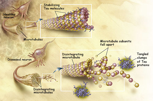
Researchers have developed a molecular compound CLR01 dubbed “molecular tweezers,” which prevents toxic proteins from aggregating or clumping together and killing brain cells in Alzheimer’s disease.

Researchers at UCLA demonstrated that the compound safely crossed the blood–brain barrier, cleared the existing amyloid-beta and tau aggregates, and also proved to be protective to the neurons’ synapses — another target of the disease — which allow cells to communicate with one another.

“This is the first demonstration that molecular tweezers work in a mammalian animal model,” Gal Bitan, an associate professor of neurology at UCLA and the senior author of the study, said.

“Importantly, no signs of toxicity were observed in the treated mice. The efficacy and toxicity results support the mechanism of this molecular tweezer and suggest these are promising compounds for developing disease-modifying therapies for Alzheimer’s disease, Parkinson’s and other disorders,” he said.

Bitan and his colleagues, including Aida Attar, first author of the study and a graduate student in Bitan’s lab, have been working with a particular molecular tweezer called CLR01.

In collaboration with scientists at the Università Cattolica in Rome, the researchers, working first in cell cultures, found that CLR01 effectively inhibited a process known as synaptotoxicity, in which clumps of toxic amyloid damage or destroy a neuron’s synapses.

Even though synapses in transgenic mice with Alzheimer’s may shut down and the mice may lose their memory, upon treatment, they form new synapses and regain their learning and memory abilities. “For humans, unfortunately, the situation is more problematic because the neurons gradually die in Alzheimer’s disease,” Bitan said.

“That’s why we must start treating as early as possible. The good news is that the molecular tweezers appear to have a high safety margin, so they may be suitable for prophylactic treatment starting long before the onset of the disease,” he said.

The report has been published in the journal Brain.

Questions Are Great Brain Boosters


Questions are great brain boosters. They can energize us to start a new behavior, or to break free and let go of an old one. That’s because questions can stimulate powerful emotions, such as curiosity or wonder, which put our brains in first gear, raring to go and learn. Some questions, that is.

A ground-breaking study by Swiss researchers published in Nature revealed that, though the neurons of the amygdala play a key part in processing fear, other areas, more specifically, the higher cortex can also play a key role in regulating the fear response and modulating new emotional learning. Thus fear does not have to debilitate our capacity to make better choices.

And, when it comes to dealing with fears, a good question can empower us to muster the courage to face challenges and fears, and perhaps discover new possibilities in the process!

Not all questions, however, energize optimal processes and our brain’s amazing capacity to learn and create new understanding in the process. Some questions achieve the opposite, and some of these aren’t ‘real’ questions.

‘Real’ versus rhetorical-why-loop questions?
Whereas most questions likely fall in low to high average range in terms of their potential for positive or negative impact on our brain’s otherwise amazing ability to change, heal and grow, arguably, all brains may be better off without the type of questions in the category of rhetorical-why loops.

The rhetorical-why-loop is not a question at all, and more like an indictment.

Whereas ‘real’ questions open space for some level of conversational exchange or possibility thinking, rhetorical-why-loops can hold a person’s mind (and body) hostage to rigid perceptions that cause them to treat losses, mistakes or failures as if they have power to permanently taint the value of a person or situation.

This position casually casts aside the possibility of restoring balance (via some level of acceptance that necessitates engaging higher cortex functions, such as reflective thinking), and automatically (subconsciously) demands harsh treatment instead, perhaps even infinite punishment – and the expectation that all concerned agree.

Examples of rhetorical-why loops include:
“Why me?”
“Why did this happen?”
“Why did this happen to me?”
“Why did you do this to me?”
Often these questions involve the use of absolute adverbs, such as always and never, for example:
“Why does this always happen?”
“Why does this only happen to me?”
“Why do you/they never listen to me?”
“Why do you/they always ignore me?”
In some cases, the questions may indict a relationship or target an idea or institution, in many cases God or Life itself. The body operating with fear in charge can act in desperate and irrational ways.
“Why, God?”
“Why would God let this happen?”
An important clarification: The above questions in and of themselves are not the problem per se.
It is quite natural to ask some or all of the above in some phases of dealing with information that pains or shocks us. And of course it is also possible to verbalize the questions in a light-hearted probing or joking way, and so on.

So what makes rhetorical-why-loops distinct? Their driving intent.

As a survival response, the intent of rhetorical-why loops seems to be to banish or attack the value, adequacy or worth of one or more human beings or life — and not to learn anything of value, discover new resources, choices or useful insights on one’s life journey.

Arguably, whether conscious or subconscious, it is the intent that either:
  • Produces emotional-physiological states in the direction of love- or fear-based emotions.
  • Decides whether the autonomic nervous system will remain in parasympathetic or activate sympathetic state.
  • Overall enriches or blocks optimal brain processes (depending on the intensity).
To be fair, there is a more “benevolent” underlying intent to keep in mind. Absent the skill or know-how to more effectively deal with or lower the intensity of painful emotions (which the body regards as a most pressing need), as protective or defensive strategies, this helps us avoid and redirect them. It also explains why we may use extreme measures (that defy logic) to distance and not feel or deal with painful emotions directly (which is not helpful in the long term!).

Regardless how benevolent, the overall effect of rhetorical-why-loops can result in a coup d’état of a person’s mind and body to where they may come to totally believe that if only they could get an answer to the ‘why’ question they ask, things would change or go back to a pervious time when the pain was not present. 
In sum, whereas rhetorical-why-loop questions can block the brain’s natural ‘learning’ mode and keep us stuck in ‘protection’ mode, real questions empower us to break this hold and find the way out of toxic loops.
The intent of a question seems to be a driving factor.

A good question can energize us to more effectively deal with fears, sustain optimal states of mind and body, courageously face challenges and fears, inquire in new directions, probe more deeply for understanding – and even create new possibilities, seemingly out of thin air!

In Part 2, how rhetorical-why-loop questions can trip us up, and what empowering questions to ask instead.

The brain man

SOME people collect stamps, others vintage cars. As a young PhD student at the University of Cambridge in the 1980s, Claude Wischik was on a mission to collect brains. 

http://www.askdrmakkar.com/images/AD_tangles.jpgIt wasn't easy. At the time, few organ banks kept entire brains. But Wischik, an Australian who was in his early 30s at the time, was trying to answer a riddle still puzzling the scientific community: what causes Alzheimer's disease?

To do that, he needed to examine brain tissue from Alzheimer's patients soon after death. That meant getting families' approval and enlisting mortuary technicians to extract the brains, he says, "no matter the time of day or night". And it wasn't just a few brains: he collected more than 300 across about a dozen years.
In his lifelong investigation, Wischik has backed a minority scientific view

that a protein called tau - which forms twisted fibres known as tangles inside the brain cells of Alzheimer's patients - is largely responsible for driving the disease.

For 20 years much of the scientific community, with billions of dollars of pharmaceutical investment, has supported a different theory that places chief blame on a different protein, beta amyloid, which forms sticky plaques in the brains of sufferers.

But a string of experimental drugs designed to attack beta amyloid has failed recently in clinical trials, and after years on the sidelines Wischik sees this as tau's big moment.

In the early 90s he and his colleagues compared the brains of Alzheimer's sufferers with those of people who died without dementia, to see how their levels of amyloid and tau differed.

They found healthy and Alzheimer's brains could be filled with amyloid plaque but only Alzheimer's brains contained aggregated tau.

What's more, as the levels of aggregated tau in a brain increased, so did the severity of dementia.
"We decided that amyloid isn't what is making people demented," Wischik says.

In the mid-90s he discovered that a drug sometimes used to treat psychosis dissolved tangles in a test tube.
He tried to set up a company to develop the drug as a treatment for Alzheimer's but found American and British venture capitalists wanted to invest in amyloid projects, not tau.

By 2002 he had scraped together $5 million from Asian investors with the help of a Singapore physician who was the father of a classmate of Wischik's son at Cambridge.

The company Wischik co-founded 10 years ago, TauRx Pharmaceuticals, is based in Singapore but conducts most of its research in Scotland, where he now lives. As his tau effort was launched, early tests of drugs designed to attack amyloid plaques were disappointing.

A vaccine developed by Athena Neurosciences failed to improve patients' cognitive function in a trial that ended in 2002.

To better understand these results, a team of British scientists largely unaffiliated with Athena or the failed clinical trial decided to examine the brains of patients who had taken part in the study. They waited for the patients to die, then, after probing the brains, concluded that the vaccine had indeed cleared amyloid plaque but had not prevented further neuro-degeneration.

In 2004 TauRx began a clinical trial of its drug, called methylene blue, in 332 Alzheimer's patients.
About the same time, a drug maker called Elan Corp, which had bought Athena Neurosciences, began a trial of an amyloid-targeted drug called bapineuzumab in 234 patients.

A key moment came in 2008 when Wischik and Elan presented results of their studies at an Alzheimer's conference in Chicago.

The Elan drug failed to improve cognition any more than a placebo pill did and Elan shares plummeted by more than 60 per cent across the next few days.

The TauRx results Wischik presented were more positive, though not unequivocal. The study showed that after 50 weeks of treatment Alzheimer's patients taking a placebo had lost 7.8 points in a test of cognitive function, while people taking 60mg of TauRx's drug three times a day had lost only one. This amounted to an 87 per cent reduction in the rate of decline for people taking the TauRx drug.

But TauRx did not publish a full set of data from the trial and this led to some scepticism among researchers. (Wischik says it didn't protect the company's commercial interests.)

What's more, a higher, 100mg dose of the drug didn't produce the same positive effects in patients. Wischik blames this on the way the 100mg dose was formulated and says the company is testing a tweaked version of the drug in its new clinical trials, which will begin enrolling patients this year.

With its new clinical trial program under way, TauRx is the first company to test a tau-targeted drug against Alzheimer's in a large human study, known in the industry as a phase 3 trial.

Wischik admits he may be just as much a zealot about tau as he accuses others of being about beta amyloid. "I may be," he says. "In the end ... it's down to the phase three trial."

Albert Einstein’s brain may provide clues to his genius

Albert
Called the “embodiment of pure intellect,” Albert Einstein has long been considered one of the most brilliant men who ever lived. During his life and since his death, people everywhere have wondered how one man could have possessed such genius. Now, scientists may have uncovered a clue within the physicist’s unusual brain. The images of Einstein’s brain are published in Falk, Lepore & Noe 2012, (The cerebral cortex of Albert Einstein: a description and preliminary analysis of unpublished photographs, “Brain”) and are reproduced here with permission from the National Museum of Health and Medicine, Silver Spring, Md.

According to a new study led by Florida State University evolutionary anthropologist Dean Falk, “portions of Einstein’s brain have been found to be unlike those of most people and could be related to his extraordinary cognitive abilities.”

“Certain things are normal,” Falk told The Huffington Post in a phone interview. “Brain size is normal. Overall shape is asymmetrical, and that is normal. What is unusual is the complexity and convolution in the various parts of the brain.”

According to a written statement issued by the university, the study, published Nov. 16 in the journal “Brain,” reveals the description of Einstein’s entire cerebral cortex. To do this, Falk and her colleagues examined 14 recently uncovered photographs of Einstein’s brain — photos that, Falk said, were difficult to obtain.

When Einstein died in 1955, his brain was removed by Thomas Harvey, a doctor at the hospital where the physicist died, NPR notes. It is likely that Harvey never got permission to remove the brain, but as author Brian Burrell writes in “Postcards from the Brain Museum,” the doctor got a posthumous stamp of approval from Einstein’s son. Harvey had said that he intended to study the brain, or at the very least, to find other scientists to do so — something that was never satisfactorily achieved in the doctor’s lifetime.

Still, scientists are now able to study Einstein’s brain thanks to a number of photos and specimen slides that Harvey had prepared of the organ. The brain, which was photographed from multiple angles, also has been sectioned into 240 blocks from which histological slides were made.

As the FSU statement notes, most of the photographs, blocks and slides were lost from public sight for more than 55 years; fortunately, a number of them have been recently rediscovered and some can now be found at the National Museum of Health and Medicine. It was with a few of these images, 14 to be exact, that Falk and her colleagues were able to take a closer look at Einstein’s brain.

What they discovered was astonishing.

“Although the overall size and asymmetrical shape of Einstein’s brain were normal, the prefrontal, somatosensory, primary motor, parietal, temporal and occipital cortices were extraordinary,” said Falk, who compared the organ to 85 other human brains already described in the scientific literature. “These may have provided the neurological underpinnings for some of his visuospatial and mathematical abilities.”

Optogenetics illuminates pathways of motivation through brain, Stanford study shows

STANFORD, Calif. — Whether you are an apple tree or an antelope, survival depends on using your energy efficiently. In a difficult or dangerous situation, the key question is whether exerting effort — sending out roots in search of nutrients in a drought or running at top speed from a predator — will be worth the energy.
http://hplusmagazine.com/sites/default/files/images/articles/optogenetics-2.jpgIn a paper to be published online Nov. 18 in Nature, Karl Deisseroth, MD, PhD, a professor of bioengineering and of psychiatry and behavioral sciences at Stanford University, and postdoctoral scholar Melissa Warden, PhD, describe how they have isolated the neurons that carry these split-second decisions to act from the higher brain to the brain stem. In doing so, they have provided insight into the causes of severe brain disorders such as depression.
In organisms as complex as humans, the neural mechanisms that help answer the question, "Is it worth my effort?" can fail, leading to debilitating mental illnesses. Major depressive disorder, for instance, which affects nearly 20 percent of people at some point in life, is correlated with underperformance in the parts of the brain involved in motivation. But researchers have struggled to work out the exact cause and effect.

"It's challenging because we do not have a fundamental understanding of the circuitry that controls this sort of behavioral pattern selection. We don't understand what the brain is doing wrong when these behaviors become dysfunctional, or even what the brain is supposed to be doing when things are working right," Deisseroth said. "This is the level of the mystery we face in this field."

Clinicians refer to this slowing down of motivation in depressed patients as "psychomotor retardation." According to Deisseroth, who is also a practicing psychiatrist, patients may experience this symptom mentally, finding it hard to envision the positive results of an action, or, he said, they may feel physically heavy, like their limbs just do not want to move.

"This is one of the most debilitating aspects of depression, and motivation to take action is something that we can model in animals. That's the exciting opportunity for us as researchers," said Deisseroth, who also holds the D.H. Chen Professorship.


Light coercion
Psychiatrists, Deisseroth included, believe the will to act may be born in the prefrontal cortex — the foremost part of the brain that helps plan and coordinate action. It then zips through the brain as a series of electrical signals, passing from neuron to neuron along countless branching pathways until it reaches the nerves that directly implement movement. Until this study, however, it was not clear which of these pathways might control the willingness to meet challenges, or the anticipation that action might be worthwhile in a difficult situation.

To isolate these pathways relevant to depression, Deisseroth's team needed to stimulate specific brain cells in rodents and observe changes in their behavior. They used optogenetics, a technique Deisseroth developed at Stanford in 2005, which has since revolutionized the fields of bioengineering and neuroscience.

The secret is as old as green algae. These single-celled organisms produce a protein called channelrhodopsin that makes them sensitive to sunlight. Borrowing and engineering the gene for this protein, Deisseroth has been able to create neurons that respond to light delivered from fiber-optic cables. He can turn the neurons on and off by sending bursts of light to activate different areas of the brain and then observe the effects on behavior.


Working backward
Surprisingly, the researchers found that simply stimulating the prefrontal cortices of rodents didn't motivate them to try any harder in a laboratory challenge. It turns out that motivation is not as simple as stimulating a region of the brain. Instead of one switch in the prefrontal cortex that turns motivation on, multiple switches work in concert. Some neurons excite motivated activity and others inhibit it. Broadly stimulating the executive part of the brain will not generate a simple effect on behavior.

"It's one step more subtle" said Deisseroth, "but this is something that optogenetics was very well-suited to resolve."

An optogenetic method called projection targeting allowed the scientists to work backward from the brain stem and find the exact pathway from neurons in the prefrontal cortex that signal motivation.

The researchers first introduced their light-sensitive protein into cells in the prefrontal cortex. The light sensitivity then spread out like the branches of a tree through all the outgoing connections and eventually made its way to the brain stem, making those regions light sensitive, too.

Then, illuminating the newly light-sensitive regions of the brain stem thought to control motivational movement, Deisseroth and Warden watched the behavioral effects as a subgroup of neurons in the prefrontal cortex that sent connections to brain stem were activated. They could see not only which cells are possibly involved in motivation, but the way motivation moves from one brain region to another.


Mapping motivation
The researchers suspected that one part of the brain stem in particular, the dorsal raphe nucleus, might be crucial to behaviors that control effort. This cluster of cells is a production hub for serotonin — a chemical messenger that changes the firing behavior of other cells. Serotonin is associated with mood modulation; many antidepressant drugs, for instance, may act by increasing serotonin concentration in the brain.

When the pathway between the prefrontal cortex and the dorsal raphe nucleus was stimulated, rodents facing a challenge in the lab showed an immediate and dramatic surge in motivation.

Curiously, however, when the rodents were relaxing in their home environment, the same stimulation had no effect. The pathway was not merely linked to any action, or to agitation; it was, more specifically, helping to "set the effort that the organism was willing to put forth to meet a challenge," Deisseroth said.

Researchers were also able to produce the opposite effect — reduced effort in response to challenge — by stimulating prefrontal neurons that project to the lateral habenula, a region perched atop the brain stem that is thought to play a role in depression. When this region was getting signals driven optogenetically from the prefrontal cortex, rodents put forward less effort.


Larger puzzles
These findings are part of a larger puzzle that Deisseroth and his team have pieced together by using optogenetics to model human behavior in animal subjects. The work has already helped clinicians and researchers to better understand what is going on in a patient's brain.

Connecting depressive symptoms with brain pathways may be helpful in the development of drugs, but according to Deisseroth, the most important part of this research is its insight into how motivation works in both depressed and healthy people.

He has observed that this insight alone can be helpful to those dealing with mental illness and seeking an explanation for troubling symptoms that feel deeply personal. For those patients, he said, simply knowing that a biological reality underlies their experience can be a motivational force in itself.

Brain on Fire

‘A relatively treatable autoimmune illness, Anti-NDMA-receptor encephalitis, a brain lobe swell detected by brain biopsy.’
http://blogs.nottingham.ac.uk/researchexchange/files/2012/06/brain-on-fire-420x210.jpg
A Manilan spending summers with relatives in the rural, I remember hearing of a young man “whose body and mind were demon-possessed, had turned deranged and unmanageable that his impoverished family had to take him to the deepest forest; left him there to live and die by himself.”   Filipinos read and hear too often of catatonic people of all ages, believed to have been turned that way by mangkukulam; possessed by witches; made fun of by middle-earth creatures called dwende;  and all sorts of frightening superstitions accepted by  many as realities.
 
A new memoir,  ‘Brain on Fire: My Month of Madness’ piece together Susannah Cahalan’s physical and mental breakdown, her lost terrifying one month in the hospital, and the grueling year it took to recover.
Sarah B. Weir,  a Yahoo! blogger  tells us the story:   That before Susannah Cahalan, 24, mysteriously contracted the disease, she was a bright, outgoing, and ambitious reporter of the New York Post.

Weir continues with her narration about Cahalan:    After exhibiting flu-like symptoms that were initially diagnosed as mono, the victim suddenly began experiencing delusions and behaving erratically. Within a few weeks, she became increasingly abusive, moody, and paranoid. Her doctors brushed off her condition as a result of too much partying and stress, but her first violent seizure signaled there was something critically askew.

Late one night, Cahalan’s guttural moans and grating squeaks woke up her boyfriend, Stephen. “My arms suddenly whipped out in front of me like, like a mummy, as my eyes rolled back and my body stiffened,” Cahalan writes. “I was gasping for air. My body continued to stiffen as I inhaled repeatedly, with no exhale. Blood and foam began to spurt through clenched teeth. Terrified, [he] stifled a panicked cry and for a second he stared, frozen, at my shaking body.”   Cahalan now describes her seizures as eerily similar to the character Regan’s outbursts in ‘The Exorcist.’

Cahalan was  plunged into a nightmare world of paranoia, psychosis, and ultimately, catatonia.  In another era, it’s likely she would have been permanently institutionalized or given a lobotomy.  In another culture, she might have been exorcised for demonic possession.  [In back of beyond locations in the Philippines, the local priest would have been called to do exorcism with a Cross and holy water to get the devil out of her body.
 That failing, the undiagnosed victim  may have been left in the jungle to fare for herself.  In Manila, she may have been confined at the National Psychopatic Hospital chained to her bed.]

Cahalan was admitted to the New York University Medical Center, and spent a month that was forever erased from her memory as her brain short-circuited. Only later, by cobbling  physicians’ notes and her father’s journal, and viewing chilling hospital videos would she fully understand the extent of her disintegration. Her frontal lobe function was almost at zero and the medical staff couldn’t be sure the right hemisphere of her brain would be salvageable. Although $1 million worth of medical tests provided few clues to her illness, her parents never gave up. “They were completely focused on finding an answer.”

Her savior, who she lovingly refers to as Dr. House, was Souhel Najjar, a Syrian immigrant.  While her other doctors had all but given up on finding a diagnosis, Dr.  Najjar swiftly ordered a brain biopsy that would confirm his hunch that she was suffering from an autoimmune disease that had been identified only two years earlier.

“[Dr. Najjar]  life’s experience shaped who he is as a doctor, and he also happens to be brilliant,” says Cahalan. “He’s so adamant about getting the full sense of you as a person…he was told that he was too slow for his elementary school. He’s made it his life’s mission to not let people fall through the cracks.”
Cahalan was the 217th person in the world to be diagnosed with anti-NDMA-receptor encephalitis, a relatively treatable illness that causes swelling in the right lobe of the brain. Untreated, she may have sunk into coma and eventually died.

Najjar also provided the title to her book. “At a pivotal moment in my disease, he pulled my parents out of the hospital room and literally said to them, ‘Her brain is on fire,’” Cahalan tells Shine. “At that point, they felt it was a relief to hear that. Describing it in layman’s terms gave them some hope.”

Cahalan wants her story to help people who might “otherwise get lost in the system.” She tells Shine [a publishing institution in the USA].   “We don’t understand how neurological autoimmune disorders work. They are so under diagnosed. About 75 percent occur in women who may get told they are just stressed. Or they are hysterical.  My disease was only discovered in 2007—how many more diseases haven’t been identified yet?”

Freestyle rapping is good for the brain

Brain


Freestyle rapping – the art of spontaneously improvising lyrics in real time - doesn't just inspire awe in fans. It apparently does wonders for the brains of those who make up lyrics on the spot.

After using functional magnetic resonance imaging to study the brain activity of rappers when they are freestyling, researchers at the U.S.-based National Institute of Health concluded that the process is similar to that of other spontaneous creative acts, including jazz improvisation.

The team, led by Dr. Siyuan Liu, scanned the brains of 12 freestyle rap artists who had at least five years of rapping experience while they performed two tasks using an identical 8-bar musical track. For the first task, they improvised rhyming lyrics and rhythmic patterns guided only by the beat. In the second task, they performed a well-rehearsed set of lyrics.

When the rappers freestyled, the researchers observed increases in brain activity in the medial prefrontal cortex, a brain region responsible for motivation of thought and action.

Vocal improvisation also increased brain activity in the perisylvian system (involved in language production) and in the amygdala (an area of the brain linked to emotion), suggesting that improvisation engages a brain network that links motivation, language, mood, and action.

The findings were published online in the Nov. 15 issue of Scientific Reports.

Drug offers brain cancer victims extra weeks of normal life

Patients with incurable brain tumours could be given new hope thanks to a drug currently used on bowel cancer, a study suggests.

Glioblastoma multiforme (GBM) kills more people under 40 than any other cancer. Each year in the UK, around 3,000 are diagnosed with the disease, the most common and most dangerous of brain tumours.

Unlike other cancers, which are more likely to strike as patients get older, GBM is just as prevalent in patients  who are young and healthy.

Unfortunately, the average sufferer will only survive for 14 months after diagnosis and 2,500 die from their tumours annually. New hope: A new trial has found Avastin may help slow the affects of brain cancer
New hope: A new trial has found Avastin may help slow the affects of brain cancer for patients

However, a new trial published yesterday shows patients can be given an extra four-and-a-half months without their condition worsening if they also receive the drug Avastin.

The trial on 911 men and women suggests Avastin can slow the growth of the tumour, giving patients a few more months of relatively normal life before the tumour grows so big that it starts to destroy their ability to speak, their behaviour, their memory and their movement.

Normally, a patient with GBM will have around six months between diagnosis and treatment, and when they relapse and their condition deteriorates. The new trial suggests Avastin could boost that to around ten months.

Dr Kirsten Hopkins, a consultant clinical oncologist at the Bristol Oncology Centre who was in charge of the UK branch of the trial, said that although the benefit might sound small, a few weeks would be extremely important for patients.

‘These patients are often young and this disease is devastating. Everyone I speak to in the medical world feels that if they were the ones diagnosed, they would want to be themselves for as long as possible,’ she says. 

‘This is a time when patients need to be able to talk to their family, do things with loved ones, discuss the future and what their wishes are for when they have passed away. 
Fast killer: Glioblastoma multiforme (GBM) - the most common and dangerous type of brain tumours - kills 2,500 every year
Fast killer: Glioblastoma multiforme (GBM) - the most common and dangerous type of brain tumours - kills 2,500 every year 
‘Giving them a few extra months to do that before they deteriorate and cannot speak is important. This is an endpoint in itself, even if this drug does not improve overall survival rates.’

The results of the AvAglio trial, presented at the Society for Neuro-Oncology annual meeting in Washington, do not reveal whether patients who took Avastin also survived for longer, but this set of data is due to be published early next year.

At the moment, patients diagnosed with GBM are usually offered surgery to remove the tumour, followed by cycles of chemotherapy and radiotherapy. For most, however, relapse is inevitable and half will have died from the disease within  14 months. Around 25 per cent will manage to survive for two years, while fewer than ten per cent live for five years.

Avastin, which is made by the pharmaceutical giant Roche, works by reducing blood supply to the tumour and slowing its growth. It is already used to treat colorectal, breast and ovarian cancers.

Some patients in the UK already receive Avastin to treat recurrent forms of brain cancer, but most have had to apply through the Cancer Drugs Fund because it is not yet approved for this use on the NHS.

Charities welcomed the  news but said they wanted to know more about any possible side-effects of taking Avastin,  as well as ensure it was given  to patients before they deteriorated. They pointed  out that at the moment only  0.7 per cent of total NHS  cancer funding is spent on  brain tumours.

Colin Speirs, founder of the charity Headcase, lost his wife Becky to GBM when she was only 40. She was diagnosed in 2009 and died 14 months later, leaving three young children.

‘In principle, anything that slows the progression of GBM has to be a good thing,’ he said.

‘But this disease is such a minefield and it’s important to remember different patients are affected differently, depending on which side of the brain the tumour is found. 

‘My wife was climbing mountains after she was diagnosed but then the tumour progressed and it was on the  left of her brain, so it affected movement, personality  and memory. 

‘I would want any new drug to ensure it gives patients four more months when they can climb mountains and not four more when the disease has already robbed them of their speech and memory.’

It currently takes the average GP three months to diagnose GBM. 

This is because symptoms include severe headaches, vomiting and blurred vision, which can be attributed to other conditions such as migraine. Sufferers may also experience an itchy head and feel as if something is running across their scalp.

Figures suggest that in the next few years, about 20,000 Britons will be diagnosed with brain tumours. 

Three in every four will be  the result of cancers in other parts of the body spreading to the brain.