Got a bad memory? The brain has a unique way of helping you forget.

Say you’re on a date and you trip and fall so your dress rides up and he sees your underwear. Or your boss tells you that for the third year in the row there will be no raises. Both of these experiences feel uncomfortable, but what do you do to forget these awkward memories? Researchers found that we use two different ways -- suppression or substitution -- to avoid thinking of uncomfortable or unhappy memories.
“We assume that, in everyday life, healthy people will use a mixture of both mechanisms to prevent an unwanted memory from coming to mind,” says Roland Benoit, a scientist at the Medical Research Council, Cognition and Brain Sciences Unit at University of Cambridge, via email. “We did not know whether the processes of direct suppression and thought substitution can be isolated, and which, if any of them, would actually cause forgetting.”
Roland and his co-author, Michael Anderson, asked 36 adults to participate in a memory exercise where half suppressed memories and the other half substituted new memories. The researchers hoped to understand how we voluntarily forget and how it affects general memory. The subjects were tested during magnetic resonance imaging procedures, or MRIs, allowing the researchers to observe how the brain works during suppression and substitution.
While both processes cause forgetting, a different region of the brain controls each one. When people suppress memories, the dorsal prefrontal cortex inhibits activation in the hippocampus, which plays an important role in retaining memories.
“It thus effectively breaks the remembering process. This, in turn, disrupts the memory representations that would be necessary for recalling the unwanted memory later on,” Benoit explains.
When it comes to substitution, the brain works a bit differently -- the caudal prefrontal cortex and midventrolateral prefrontal cortex form a network of sorts that works with the hippocampus to swap out new information with details people would soon forget.
“By just looking at how well people forgot memories, you couldn’t tell whether they had done direct suppression or thought substitution,” Benoit says. “These mechanisms are based on different brain systems that work in opposite fashion: One (direct suppression) by ‘slamming the mental break’ to stop the remembering process and the other (thought substitution) by steering the remembering process towards a substitute memory.”
Even though people exploit both to forget those nagging, unwanted memories, actively overlooking unpleasant events can negatively impact how we remember. But Benoit notes that learning how people deal with unwanted memories helps them understand how people with traumatic memories, such as PTSD sufferers, cope with remembering.
“It is perfectly natural for people, upon encountering an unwelcome reminder, to try to put the unpleasant reminding out of mind. We all have experienced this. Intuitively, it feels as though we solved this problem.”
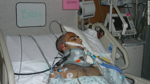 Bobby Ghassemi was just 17 years old when he was in a horrific car accident.
 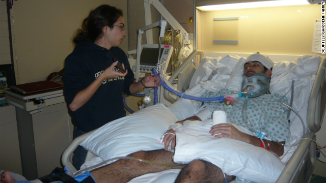 After the accident
in March 2010, doctors told Bobby's family that he could live out the
rest of his life in a vegetative state.
After the accident
in March 2010, doctors told Bobby's family that he could live out the
rest of his life in a vegetative state. 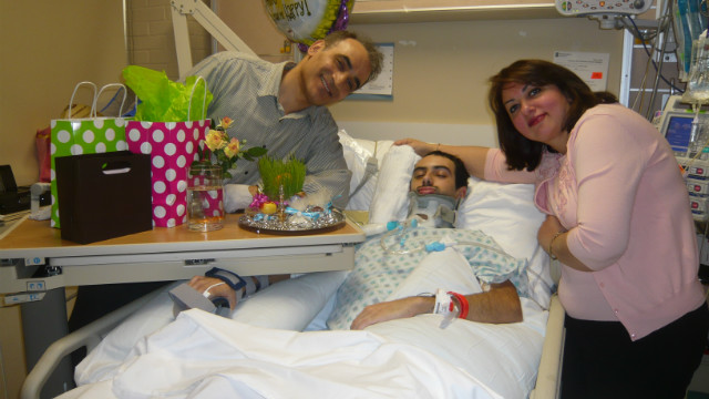 Ten days after the
accident, Bobby was still in a coma. Bobby's father, Peter, talked with
Dr. Michael Lewis, who suggested that omega-3 fatty acids might be able
to help. Peter insisted that his son be given high doses of fish oil
through a feeding tube.
Ten days after the
accident, Bobby was still in a coma. Bobby's father, Peter, talked with
Dr. Michael Lewis, who suggested that omega-3 fatty acids might be able
to help. Peter insisted that his son be given high doses of fish oil
through a feeding tube. 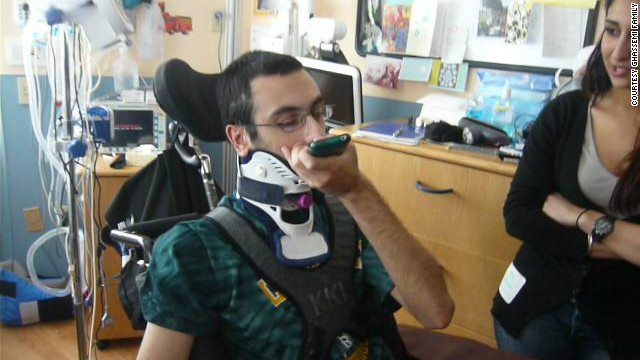 Bobby says the omega-3 fatty acids have helped in recovering his motor skills.
 .
According to a 2010 study in CA: A Cancer Journal for Clinicians, the
average survival rate after a glioblastoma diagnosis is 14 months
(though improving surgical techniques had boosted that number from 10
months in only five years prior to the study).
.
According to a 2010 study in CA: A Cancer Journal for Clinicians, the
average survival rate after a glioblastoma diagnosis is 14 months
(though improving surgical techniques had boosted that number from 10
months in only five years prior to the study).

